Details of DPV and References
DPV NO: 7 June 1970
Family: Tombusviridae
Genus: Carmovirus
Species: Carnation mottle virus | Acronym: CarMV
Carnation mottle virus
M. Hollings Glasshouse Crops Research Institute, Littlehampton, Sussex, England
Olwen M. Stone Glasshouse Crops Research Institute, Littlehampton, Sussex, England
Contents
- Introduction
- Main Diseases
- Geographical Distribution
- Host Range and Symptomatology
- Strains
- Transmission by Vectors
- Transmission through Seed
- Transmission by Grafting
- Transmission by Dodder
- Serology
- Nucleic Acid Hybridization
- Relationships
- Stability in Sap
- Purification
- Properties of Particles
- Particle Structure
- Particle Composition
- Properties of Infective Nucleic Acid
- Molecular Structure
- Genome Properties
- Satellite
- Relations with Cells and Tissues
- Ecology and Control
- Notes
- Acknowledgements
- Figures
- References
Introduction
-
Described by
Kassanis (1955).
Synonym:
- None; carnation mosaic virus is not an acceptable name because it has been applied to many different viruses
-
An RNA-containing virus with isometric particles about 28 nm in diameter, transmissible by inoculation of sap unless prevented by inhibitors. It occurs naturally only in the Caryophyllaceae but can be transmitted experimentally to other species. Very common and widespread in carnation, many commercial cultivars being wholly infected. Vector unknown.
Main Diseases
Mild mottle and loss of vigour in carnation. Symptoms often apparent only by comparison with healthy plants.
Geographical Distribution
Widespread wherever carnations are grown.
Host Range and Symptomatology
Natural host range seems restricted to species of Caryophyllaceae, but the virus can infect over 30 spp. in 15 dicotyledonous families. Because of inhibitors in carnation sap the virus may be transmitted readily only after purification.
-
Diagnostic species
- Chenopodium amaranticolor.
Usually develops local chlorotic dots after 4-7 days (Fig. 1); lesions may be necrotic with some strains or in older leaves. Systemic infection rare, and usually occurs only in short day conditions. - Chenopodium quinoa. Local chlorotic spots after 4-7 days followed by systemic
chlorotic spotting and mottle with some leaf distortion.
- Atriplex hortensis. Local chlorotic spots after 5-9 days followed by systemic chlorotic flecks and leaf buckling.
- Gomphrena globosa. Faint fawn local necrotic dots after 7-10 days (Fig. 5). No systemic infection; this distinguishes the virus from carnation ringspot virus.
- Tetragonia expansa. Faint local chlorotic or necrotic spots after 5-8 days (Fig. 7) followed by limited systemic infection.
- Dianthus barbatus (sweet william). Some clones give local fawn spots after 4-7 days (Fig. 4) followed by systemic chlorotic flecking and mottling; others are immune.
- Atriplex hortensis. Local chlorotic spots after 5-9 days followed by systemic chlorotic flecks and leaf buckling.
-
Propagation species
- Dianthus caryophyllus
(carnation) is very suitable both for maintaining the virus and as a source of virus for purification. Chenopodium quinoa is also useful; Dianthus barbatus should only be used where a susceptible host clone has been selected.Assay species
- Chenopodium amaranticolor
and C. quinoa are useful local lesion hosts and will detect attenuated forms of the virus (see below).
Strains
Three minor variants are recorded:
PSR strain (Hollings & Stone, 1964). Produces necrotic local lesions in a number of hosts which give chlorotic spots with the type strain (Fig. 2, Fig. 7). It is serologically indistinguishable from the type strain.
PR4 strain (Kemp & Fazekas, 1966). Can be distinguished from the type strain serologically and by the smaller local necrotic lesions produced in Dianthus barbatus.
Attenuated form (Hollings & Stone, 1962). Found in carnation plants after heat treatment or meristem tip culture. Very few, large local chlorotic lesions develop in Chenopodium amaranticolor after about 21 days (Fig. 3). On serial passage, such lesions give the typical, rapidly multiplying form with no serological difference from the type strain.
Transmission by Vectors
None found.
Transmission through Seed
None found.
Transmission by Dodder
None detected.
Serology
The virus is a good immunogen and rabbits given one intravenous and two intramuscular injections produce antisera with specific precipitin tube titres of 1/4096 to 1/16,384. Specific precipitates in tube tests are granular (somatic), and a single band of precipitate forms in gel-diffusion tests. All strains of the virus, except the attenuated form, can be detected in crude sap by gel-diffusion tests. In gel immunoelectrophoresis the virus moves towards the cathode (Devergne & Cardin, 1967).
Relationships
No relationship has been shown to any other isometric virus. The type strain and the PSR strain protected against each other in cross-protection tests.
Stability in Sap
In carnation sap the thermal inactivation point (10 min) is about 90°C though much infectivity is lost at 65°C; dilution end-point is about 10-5. Infectivity survives for 70 days at 20°C; the attenuated form was recovered after 13 months. Virus lyophilized in sap of Dianthus barbatus, stored at room temperature under vacuum, retained infectivity for over 10 years.
Purification
The method used by Hollings & Stone (1965) for carnation ringspot virus is also suitable for carnation mottle virus. Leaves minced with 0.05 M phosphate buffer, pH 7.6, containing 0.1% thioglycollic acid (w/v= 1/1.25) are squeezed through cheesecloth and the slurry treated with n-butanol to 8.5% of the final volume. The mixture is stored overnight at 2°C and the virus then separated from the aqueous phase by differential centrifugation. Preparations made with borate buffers have about half the infectivity and serological activity of those prepared with phosphate buffer. Precipitation with ammonium sulphate (40% saturation) or ethanol (50%) or by acidification to pH 4.8 also gives acceptable virus preparations.
Properties of Particles
Sedimentation coefficient (s20, w): about 122 S.
No accessory viral components are found by analytical centrifugation.
Molecular weight: 7-8 x 10⁶.
Isoelectric point: pH 5.2.
Electrophoretic mobility: -8 x 10⁻⁵ cm2 sec-1 volt-1 at pH 7 in 0.02 ionic strength buffer (Tremaine, unpublished).
Particle Structure
Particles are rounded, isometric and about 28 nm in diameter (Fig. 6). Subunit structure is not known.
Particle Composition
RNA: Single stranded, about 20% of particle weight. Molar percentages of nucleotides: G27; A30; C19; U24 (Tremaine, unpublished).
Protein: About 80% of particle weight, probably about 240 subunits per particle (Tremaine, unpublished). Subunits stated to contain 382 amino acid residues (Tremaine & Goldsack, 1968).
Relations with Cells and Tissues
The virus is found in stems, leaves, flowers and roots, though in lower concentration in apical meristems. No reported inclusion bodies, or association with cell organelles. There is no information on sites of synthesis.
Notes
Carnation mottle virus can be eliminated from carnation and Dianthus barbatus by meristem tip culture with or without previous heat treatment. It often occurs together with other isometric viruses in carnation. Carnation ringspot virus usually induces much more damaging effects in carnation, and produces conspicuous local necrotic concentric rings in Gomphrena globosa and local necrotic rings and ringspots in D. barbatus. Carnation Italian ringspot virus is distinguished by its systemic invasion of Chenopodium amaranticolor and by its serological reactions; it is a serotype of tomato bushy stunt virus. Two other small isometric viruses have been isolated from carnation but they have not yet been fully characterized; they do not induce symptoms in any of the diagnostic hosts of carnation mottle virus (Hollings & Stone, 1968).
Acknowledgements
Photographs: M. Hollings and Glasshouse Crops Research Institute.
Figures
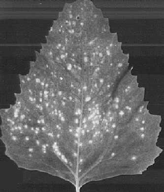
Typical local chlorotic lesions in Chenopodium amaranticolor.

Local necrotic lesions of the PSR strain in C. amaranticolor.

Local chlorotic lesions of the attenuated form in C. amaranticolor.

Necrotic local lesions in a susceptible clone of Dianthus barbatus.

Faint local necrotic lesions in Gomphrena globosa.

Virus particles from a purified preparation stained with phosphotungstate. Bar represents 100 nm.

Local necrotic lesions of the PSR strain in Tetragonia expansa.
References list for DPV: Carnation mottle virus (7)
- Devergne & Cardin, Annls. Épiphyt. 18: 65, 1967.
- Hollings & Stone, Rep. Glasshouse Crops Res. Inst., 1961: 100, 1962.
- Hollings & Stone Ann. appl. Biol. 53: 103, 1964.
- Hollings & Stone, Ann. appl. Biol. 56: 73, 1965.
- Hollings & Stone, Rep. Glasshouse Crops Res. Inst., 1967: 94, 1968.
- Kassanis, Ann. appl. Biol. 43: 103, 1955.
- Kemp & Fazekas, Can. J. Bot. 44: 1261, 1966.
- Tremaine & Goldsack, Virology 35: 227, 1968.