Details of DPV and References
DPV NO: 35 October 1970
Family: Rhabdoviridae
Genus: Nucleorhabdovirus
Species: Potato yellow dwarf virus | Acronym: PYDV
Potato yellow dwarf virus
L. M. Black Dept. of Botany, University of Illinois, Urbana, Illinois 61801, USA
Contents
- Introduction
- Main Diseases
- Geographical Distribution
- Host Range and Symptomatology
- Strains
- Transmission by Vectors
- Transmission through Seed
- Transmission by Grafting
- Transmission by Dodder
- Serology
- Nucleic Acid Hybridization
- Relationships
- Stability in Sap
- Purification
- Properties of Particles
- Particle Structure
- Particle Composition
- Properties of Infective Nucleic Acid
- Molecular Structure
- Genome Properties
- Satellite
- Relations with Cells and Tissues
- Ecology and Control
- Notes
- Acknowledgements
- Figures
- References
Introduction
-
Disease described by Barrus & Chupp (1922).
There are two closely related but distinct forms of potato yellow dwarf virus, one transmitted by Aceratagallia sanguinolenta but not by Agallia constricta (sanguinolenta yellow dwarf virus; SYDV) and one with the inverse vector relationships (constricta yellow dwarf virus; CYDV) (Black, 1941). This description is for SYDV except where stated otherwise.
Selected synonyms
- Aureogenus vastans
(Rev. appl. Mycol. 23: 490) - SYDV has been called A. vastans var. vulgare (Rev. appl. Mycol. 23: 490) and New York potato yellow dwarf virus (Rev. appl. Mycol. 21: 36). CYDV has been called A. vastans var. agalliae (Rev. appl. Mycol. 28: 514) and New Jersey potato yellow dwarf virus (Rev. appl. Mycol. 21: 36). Consult CMI Phytopath. Paper 9, 204 pp., 1968, for other synonyms
-
A virus with bacilliform particles (380 x 75 nm) containing lipid, protein and RNA. Transmitted in the persistent manner by agallian leafhoppers (in which it multiplies) and by manual inoculation to Nicotiana rustica. Host range wide; found sporadically in potato in North America.
Main Diseases
Yellow dwarf of potato.
Geographical Distribution
Virus spreads from plant to plant in northeastern USA and adjacent parts of Canada. Elsewhere it develops in potato plants grown from infected tubers. CYDV has been found only in New Jersey, but probably has a wider distribution.
Host Range and Symptomatology
All known plant hosts are dicotyledons. Nicotiana rustica and N. glutinosa can be infected by leaf abrasion inoculations, N. rustica being much the more susceptible, but, in addition to solanaceous plants, vectors have transmitted the virus to species in the families Compositae, Cruciferae, Labiatae, Leguminosae, Polygonaceae and Scrophulariaceae (Younkin, 1942; Hansing, 1942).
Varieties of Solanum tuberosum show economically important differences in disease incidence in the field (Taylor, 1938; Hansing, 1943; Larson, 1945). Medicago sativa cv. Grimm is immune (Black, 1943a). The ox-eye daisy, Chrysanthemum leucanthemum var. pinnatifidum is the principal source of virus for infecting potato crops (Younkin, 1943). Clover, the preferred host of the vector (Watkins, 1941), is rarely infected (Younkin, 1943). High temperatures favour the development of the disease in potato and low temperatures retard it (Walker & Larson, 1939). Severe winters may greatly reduce the overwintering population of clover leafhoppers. Drought forces migration of vectors into potato fields. The virus survives the winter in infected ox-eye daisies, potato tubers and adult leafhoppers.
-
Diagnostic species
- Solanum tuberosum
(potato). Apical yellowing and necrosis, internal necrotic spots in stems, particularly in upper nodes. Dwarfing or killing of sprouts before emergence. Internal necrotic spots in tubers; malformation, cracking, dwarfing of tubers. Plant rarely survives; when it does, chronic symptoms are milder. -
Nicotiana rustica. SYDV causes yellow primary lesions (Fig. 2) first detectable in about 1 week in suitable leaves inoculated by rubbing. Systemic symptoms are mottling and yellowing of leaves (Fig. 1), sometimes with light green or yellow streaking of stems. Chronic symptoms are milder. CYDV causes primary lesions of variable appearance and is more slow to infect plants systemically (Fig. 1).
Trifolium incarnatum. May be inoculated by vectors or by pricking inoculum into the crown of the plant. With SYDV, vein-clearing appears first in younger leaves (Fig. 6) and plant usually dies. With CYDV, systemic symptoms appear first in older leaves and rusty-brown spots and lines (Fig. 6) develop which never result from SYDV infection; plant usually survives acute infection by CYDV and chronic infection is very mild.
-
Propagation species
- Nicotiana rustica.
Assay species
- Nicotiana rustica
leaves of suitable age. -
Monolayers of vector cells. Cells from A. sanguinolenta are designated AS cells, those from A. constricta, AC cells. Infected cells are countable 2 days after inoculation if stained by specific fluorescent antiserum (Chiu et al., 1970). Nevertheless, no serological reactions in tissues of viruliferous vectors have been demonstrated. The infectivity of SYDV for AC cells is about l0% that for AS cells, that of CYDV for AS cells about 10% of that for AC cells (Liu & Black, 1969).
Strains
Strains of differing virulence may be isolated from primary lesions of SYDV (Black, 1940). Strains that are transmitted poorly or not at all by the vector arise when the virus is maintained in N. rustica for long periods without passage through the vector (Black, 1953b, 1969).
Transmission by Vectors
Crimson clover is the best host for testing vectors. SYDV is transmitted well by Aceratagallia sanguinolenta (Fig. 3) (Black, 1934), and several other Aceratagallia spp., poorly by Agallia quadripunctata and not at all by Agallia constricta. CYDV is transmitted by Agallia constricta but not by Aceratagallia sanguinolenta. Agallia quadripunctata transmits CYDV somewhat better than SYDV. The viruses multiply in the vectors (Chiu et al., 1970). Nymph, adult male and adult female insects transmit. The virus can overwinter in adult vectors even in the absence of food plants (Black, 1937). Minimum incubation period in the vector is 6 days. In some tests SYDV (Black, 1943b) and CYDV (Nagaraj & Black, 1962) were not transmitted through egg or sperm of the vector but in others CYDV was occasionally transmitted through the egg (Black, 1953a). Individuals of A. sanguinolenta and A. constricta differ genetically in ability to transmit SYDV and CYDV respectively (Black, 1943b; Nagaraj & Black, 1962). Sinha (1965) recovered infective CYDV from various organs of A. constricta.
Transmission through Seed
No seed transmission found in N. rustica.
Transmission by Dodder
No information.
Serology
Rabbit antisera with titres of 1/1280 can be produced by intramuscular injection of purified virus in adjuvant. Antisera react with virus and soluble antigen from plants but neither reaction has been demonstrated with extracts from vectors (Wolcyrz & Black, 1956).
Relationships
Tests with virions or soluble antigen show that SYDV and CYDV are serologically related but not identical (Wolcyrz & Black, 1957). CYDV and strains of SYDV protect N. rustica from severe infection with a necrotizing strain of SYDV whereas several unrelated viruses do not (Black, 1940, 1944).
Stability in Sap
In sap of N. rustica, infectivity is retained at 23-27°C for 2½-12 hr, dilution end-point is usually about 10-3, thermal inactivation point (10 min) about 50°C. Virus does not withstand desiccation in N. rustica leaves or prolonged storage in frozen leaves.
Purification
Purified by density-gradient zonal centrifugation and electrophoresis (Brakke, 1951, 1953, 1955). Certain solutions, e.g. 0.1 M glycine and 0.01 M MgCl2, noticeably stabilize infectivity (Brakke, 1956). Later methods are in essence modifications of Brakke’s techniques.
Properties of Particles
The particles seem to be of one type. The dilution curve indicates that one particle initiates infection (Chiu et al., 1970). Sedimentation coefficient (s20,w): 810-950 S.
Buoyant density (in sucrose): 1.17 (Brakke, 1958).
Molecular weight (calculated from dimensions and density): c. 1100 x 106 daltons.
Purified virus does not give a peak in its light absorption spectrum at any wavelength between 220 and 320 nm.
Particle Structure
Bacilliform particles (Fig. 4), c. 380 x 75 nm in plant sections, but smaller in sections of vector cell monolayers. Viral envelope appears to consist of three lamellae c. 3.5 nm thick and c. 5 nm apart (MacLeod, Black & Moyer, 1966). Electron micrographs of the particles of this virus taken at different times (Black, Mosley & Wyckoff, 1948; Brakke, Black & Wyckoff, 1951; Brakke & Vatter (in Black, 1955); Black et al., 1965; Macleod et al., 1966; MacLeod, 1968) illustrate various properties of the particles.
Particle Composition
The virus contains more than 20% lipid (Ahmed et al., 1964). It contains at least three different proteins and single-stranded RNA with a M.Wt of c. 4.3 x 10⁶ daltons (R. MacLeod, personal communication).
Relations with Cells and Tissues
Fluorescein-conjugated antibody studies show that antigen is at first confined to the nucleus of cells in culture. Both in the plant host and in the vector the principal site of virus assembly appears to be at the inner nuclear membrane and the principal site of virus accumulation is between the two nuclear membranes (Fig. 5). However, intranuclear assembly also occurs, and in later stages virus is found in the cytoplasm (MacLeod et al., 1966; MacLeod, 1968; Chiu et al., 1970).
Notes
SYDV and CYDV are the only leafhopper-borne viruses that are manually transmissible and produce primary lesions in inoculated leaves (Black, 1938). Their particles are morphologically similar to those of some other leafhopper and aphid-borne viruses (such as maize mosaic, Russian winter wheat mosaic, lettuce necrotic yellows and sowthistle yellow vein viruses) and also to those of some viruses of animals (such as bovine vesicular stomatitis virus) but there is no other evidence to show how they may be related.
Figures

Nicotiana rustica leaves, (left to right) healthy, systemically infected with SYDV and with CYDV.

Primary lesions of SYDV in N. rustica.
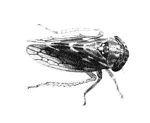
Aceratagallia sanguinolenta.

Section of N. rustica leaf showing cross and longitudinal sections of SYDV particles. Bar represents 200 nm. (Courtesy R. MacLeod.)

Section of N. rustica leaf showing accumulation of SYDV particles between separated inner and outer nuclear membranes. Bar represents 400 nm.

Trifolium incarnatum, (left) healthy, (centre) infected with SYDV, (right) infected with CYDV.
References list for DPV: Potato yellow dwarf virus (35)
- Ahmed, Black, Perkins, Walker & Kummerow, Biochem. biophys. Res. Commun. 17: 103, 1964.
- Barrus & Chupp, Phytopathology 12: 123, 1922.
- Black, Am. Potato J. 11: 148, 1934.
- Black, Mem. Cornell Univ. agric. Exp. Stn. 209: 1, 1937.
- Black, Phytopathology 28: 863, 1938.
- Black, Am. J. Bot. 27: 386, 1940.
- Black, Am. Potato J. 18: 231, 1941.
- Black, Phytopathology 33: 363, 1943a.
- Black, Genetics, Princeton 28: 200, 1943b.
- Black, Proc. Am. phil. Soc. 88: 132, 1944.
- Black, Phytopathology 43: 9, 1953a.
- Black, Phytopathology 43: 466, 1953b.
- Black, Phytopathology 45: 208, 1955.
- Black, A. Rev. Phytopath. 7: 73, 1969.
- Black, Mosley & Wyckoff, Biochim. biophys. Acta 2: 121, 1948.
- Black, Smith, Hills & Markham, Virology 27: 446, 1965.
- Brakke, J. Am. chem. Soc. 73: 1847, 1951.
- Brakke, Archs Biochem. Biophys. 45: 275, 1953.
- Brakke, Archs Biochem. Biophys. 55: 175, 1955.
- Brakke, Virology 2: 463, 1956.
- Brakke, Virology 6: 96, 1958.
- Brakke, Black & Wyckoff, Am. J. Bot. 38: 332, 1951.
- Chiu, Liu, MacLeod & Black, Virology 40: 387, 1970.
- Hansing, Phytopathology 32: 7, 1942.
- Hansing, Bull. Cornell Univ. agric. Exp. Stn 792, 28 pp., 1943.
- Larson, J. agric. Res. 71: 441, 1945.
- Liu & Black, Phytopathology 59: 1038, 1969.
- MacLeod, Virology 34: 771, 1968.
- MacLeod, Black & Moyer, Virology 29: 540, 1966.
- Nagaraj & Black, Virology 16: 152, 1962.
- Sinha, Virology 27: 118, 1965.
- Taylor, Am. Potato J. 15: 37, 1938.
- Walker & Larson, J. agric. Res. 59: 259, 1939.
- Watkins, Bull. Cornell Univ. agric. Exp. Stn 758, 24 pp., 1941.
- Wolcyrz & Black, Phytopathology 46: 32, 1956.
- Wolcyrz & Black, Phytopathology 47: 38, 1957.
- Younkin, Am. Potato J. 19: 6, 1942.
- Younkin, Fm Res. 9: 6, 1943.