Details of DPV and References
DPV NO: 75 October 1971
Family: Betaflexiviridae
Genus: Carlavirus
Species: Poplar mosaic virus | Acronym: PopMV
Poplar mosaic virus
P. G. Biddle Commonwealth Forestry Institute, Oxford, England
T. W. Tinsley Commonwealth Forestry Institute, Oxford, England
Contents
- Introduction
- Main Diseases
- Geographical Distribution
- Host Range and Symptomatology
- Strains
- Transmission by Vectors
- Transmission through Seed
- Transmission by Grafting
- Transmission by Dodder
- Serology
- Nucleic Acid Hybridization
- Relationships
- Stability in Sap
- Purification
- Properties of Particles
- Particle Structure
- Particle Composition
- Properties of Infective Nucleic Acid
- Molecular Structure
- Genome Properties
- Satellite
- Relations with Cells and Tissues
- Ecology and Control
- Notes
- Acknowledgements
- Figures
- References
Introduction
- Described by Atanasoff (1935) and Berg (1964).
- Synonym
- Canadian poplar mosaic (Rev. appl. Mycol. 14: 462)
- A virus with flexuous filamentous particles c. 675 nm long. Only known natural host is poplar, in which it is common and can cause severe growth losses. Many other plant species can be infected by inoculation of sap. There is no known vector, but it is widely disseminated in infected cuttings.
Main Diseases
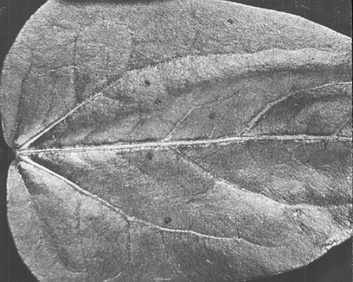
Causes a mosaic (Fig. 1) or diffuse spotting (Fig. 2) in mature leaves of most poplar clones, but some clones react more severely, with necrosis of veins and leaf stalks, swellings round the bases of the petioles, and small lesions and splits on the stem. Growth of infected trees, particularly of those showing more severe leaf symptoms, is reduced, and there are also effects on the specific gravity and strength of wood from infected trees (Biddle & Tinsley, 1971b).
Geographical Distribution
Throughout the geographical range of susceptible poplar species and clones.
Host Range and Symptomatology
Natural hosts are poplars in the Sections Aigeiros and Tacamahaca, but species in 20 dicotyledonous families can be infected by inoculation of sap (Schmelzer, 1966).
- Diagnostic species
- Nicotiana megalosiphon
(Fig. 3) and N. glutinosa. Faint chlorotic local lesions after 10 days, followed by systemic vein-clearing and leaf-curling. Veins occasionally become necrotic. - Lavatera trimestris. Small necrotic local lesions, followed by systemic
vein-clearing and vein-necrosis.
- Vigna sinensis (cowpea). Red local lesions develop after 7 days, tending to spread along the veins they contact (Fig. 4); systemic vein-clearing after about 20 days followed by vein-reddening and leaf deformation.
- Vigna sinensis (cowpea). Red local lesions develop after 7 days, tending to spread along the veins they contact (Fig. 4); systemic vein-clearing after about 20 days followed by vein-reddening and leaf deformation.
- Propagation species
- Nicotiana megalosiphon
is the most suitable species for maintaining the virus and providing material for purification.- Assay species
- Vigna sinensis
sometimes produces countable lesions, but is not a reliable assay host.
Strains
Most isolates seem identical, although Blattný (1965) distinguished a more severe strain in Populus x euramericana cv. Robusta.
Transmission by Vectors
A low rate of natural transmission has been noted in the field, but no vector has been satisfactorily implicated. Boyer (1962) claimed that the aphid Neothomasia populicola transmits the virus, but other workers have been unable to confirm this. Circumstantial evidence suggests that the virus may spread through root grafts, but from experiments with radioactive tracers, the number of root grafts seems too few to account for the rate of virus spread (P. G. Biddle, unpublished).
Transmission through Seed
Not reported.
Transmission by Dodder
Cuscuta californica and C. subinclusa failed to transmit the virus from Nicotiana glutinosa to N. megalosiphon (Schmelzer, 1966).
Serology
The virus is moderately immunogenic. Antisera with titres of 1/2048 are commonly obtained. Gel-diffusion tests are unsatisfactory, but microprecipitation tests give good results (Berg, 1964).
Relationships
The morphology of its particles and their stability and sedimentation coefficient suggest that poplar mosaic virus is a member of the potato virus S group of viruses (Brandes & Wetter, 1959). However, no interaction was detected between poplar mosaic virus and antisera against viruses of this group such as pea streak, red clover vein mosaic, carnation latent, potato S, potato M, chrysanthemum B and freesia mosaic viruses, or against other viruses with filamentous particles such as potato X, potato Y, bean yellow mosaic, iris mosaic and tulip breaking viruses (Berg, 1964).
Stability in Sap
In Nicotiana megalosiphon sap, the thermal inactivation point is about 74°C, dilution end-point about 10-5, and infectivity persists in vitro for 2 days at room temperature and for 6 days at 4°C (Schmelzer, 1966). An isolate studied by Biddle & Tinsley (1971a) was somewhat less stable and less concentrated in sap. Infectivity in poplar extracts is greatly increased by adding 0.1 g of insoluble polyvinyl pyrrolidone to each 1 ml of extraction medium.
Purification
Freeze infected Nicotiana megalosiphon leaves and triturate in buffer (0.05 M Na2HPO4, 1 mM di-sodium ethylene diaminetetraacetate, 0.2 M Na2SO3 ; pH 7.5). Heat the extract to 50°C, centrifuge at low speed, precipitate the virus by adding ammonium sulphate to 30-45% saturation, and resuspend in buffer. Further purify by centrifuging in sucrose density gradients or by clarifying with chloroform followed by molecular exclusion chromatography in columns of 2% agarose beads. The virus is liable to aggregate, and virus sedimented in the ultracentrifuge is difficult to resuspend (Biddle & Tinsley, 1971a).
Properties of Particles
Sedimentation coefficient (s20,w) at infinite dilution: 165 S (Berg, 1964).
Particle Structure
Slightly flexuous filamentous particles c. 675 nm long (Fig. 5).
Particle Composition
No reports.
Relations with Cells and Tissues
All tissues in poplar are infected, including cambium, phloem and xylem (Berg, 1964). Metabolic disturbances to the differentiating xylem result in reduced growth and abnormalities of the wood structure (Biddle & Tinsley, 1971b).
Notes
This is the only known virus of poplar. Its widespread occurrence undoubtedly arises from dissemination of infected poplar cuttings. The use of healthy plants for propagation, and rogueing of infected plants. effectively controls the disease, because natural transmission appears to be rare.
Figures
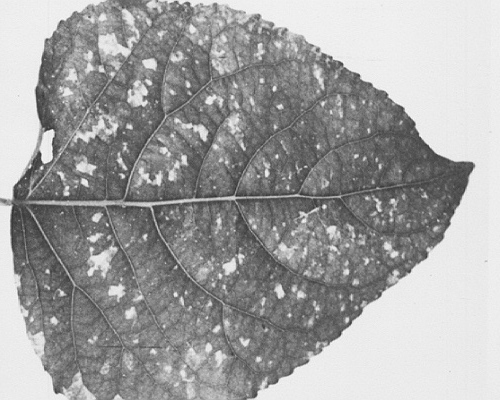
Leaf of Populus x euramericana cv. Robusta showing mosaic.

Chlorotic spots in Populus x euramericana cv. Regenerata.

Systemic symptoms in Nicotania megalosiphon.
Local lesions in Vigna sinensis.

Virus particles from clarified sap in uranyl acetate. Bar represents 100 nm.
References list for DPV: Poplar mosaic virus (75)
- Atanasoff, Phytopath. Z. 8: 197, 1935.
- Berg, Meded. LandbHoogesch. Wageningen 64: 11, 1964.
- Biddle & Tinsley, New Phytol. 70: 61, 1971a.
- Biddle & Tinsley, New Phytol. 70: 67,1971b.
- Blattný, Lesn. Cas. 11: 637, 1965.
- Boyer, Can. J. Bot. 40: 1237, 1962.
- Brandes & Wetter, Virology 8: 99, 1959.
- Schmelzer, Phytopath. Z. 55: 317, 1966.