Details of DPV and References
DPV NO: 87 June 1972
Family: Betaflexiviridae
Genus: Carlavirus
Species: Potato virus M | Acronym: PVM
Potato virus M
C. Wetter Botanisches Institut, Universität des Saarlandes, 66 Saarbrücken, Germany
Contents
- Introduction
- Main Diseases
- Geographical Distribution
- Host Range and Symptomatology
- Strains
- Transmission by Vectors
- Transmission through Seed
- Transmission by Grafting
- Transmission by Dodder
- Serology
- Nucleic Acid Hybridization
- Relationships
- Stability in Sap
- Purification
- Properties of Particles
- Particle Structure
- Particle Composition
- Properties of Infective Nucleic Acid
- Molecular Structure
- Genome Properties
- Satellite
- Relations with Cells and Tissues
- Ecology and Control
- Notes
- Acknowledgements
- Figures
- References
Introduction
- Described by Schultz & Folsom (1923) and Bagnall, Larson & Walker
(1956).
- Selected synonyms
- Kartoffel-K-Virus (Rev. appl. Mycol. 20: 486)
- Kartoffel-Rollmosaik-Virus (Rev. appl. Mycol. 15: 246)
- Potato interveinal mosaic virus (American) (McKay & Dykstra, 1932)
- Potato leaf rolling mosaic virus (Rev. appl. Mycol. 3: 548)
- Potato paracrinkle virus (Rev. appl. Mycol. 9: 604)
- Potato virus E (Rev. appl. Mycol. 12: 776; 18: 337)
- Solanum virus 7 (Rev. appl. Mycol. 36: 303)
- Solanum virus 11 (Rev. appl. Mycol. 36: 303)
- Kartoffel-Rollmosaik-Virus (Rev. appl. Mycol. 15: 246)
- A virus with straight to slightly curved filamentous particles c. 650 x 12 nm. It is sap-transmissible to a limited range of species. Most isolates are transmitted by aphids in the non-persistent manner. World-wide distribution.
Main Diseases
In potato, symptoms range from very slight (e.g. cv. King Edward) to severe (e.g. cv. Arran Victory), depending on virus strain and potato variety. Causes various mottle, mosaic, crinkling and rolling symptoms in leaves, and stunting of shoots (Fig. 1).
Geographical Distribution
World-wide in cultivated varieties of potato.
Host Range and Symptomatology
Host range is narrow. Susceptible species belong mainly to the Solanaceae. Transmissible by inoculation with sap from young leaves, but not from older leaves. May be transmitted from potato to potato by grafting.
- Diagnostic species
- Datura metel.
Chlorotic or necrotic local lesions in inoculated leaves (Fig. 2). Systemically infected leaves show rugosity and chlorotic spotting. First the lower leaves, later the upper ones are shed, and the plants become stunted and sometimes die. - Gomphrena globosa. Some isolates from mainland Europe cause local
chlorotic spots surrounded by reddish borders (Fig. 3).
- Lycopersicon esculentum (tomato). Systemically infected, but symptomless.
- Nicotiana debneyi. Irregular brown necrotic ring-like local lesions (Fig. 4). Not systemic.
- Solanum rostratum. Necrotic streak of stem, petioles and leaf-veins; systemic.
- Lycopersicon esculentum (tomato). Systemically infected, but symptomless.
- Propagation species
- Lycopersicon esculentum.
Young leaves are a suitable source of virus for purification. - Solanum tuberosum. cv. Saco may be used to maintain the virus.
- Assay species
- Datura metel
and Gomphrena globosa are local lesion hosts. When potato sap is used as inoculum, the number of local lesions on both hosts increases if the sap is heated at 50°C for 10 min. - Phaseolus vulgaris (Red Kidney bean) is reportedly a useful local lesion host (Hiruki, 1970).
Strains
Several variants can be distinguished by the symptoms they induce in potato and by slightly differing symptoms in test plants. The best known variants are:
Leaf rolling mosaic and interveinal mosaic isolates (Schultz & Folsom, 1923; Bagnall et al., 1956).
Paracrinkle isolate from potato cv. King Edward (Salaman & Le Pelley, 1930; Kassanis, 1961).
D 1102 and Fortuna isolates (Köhler, 1953; Wetter & Brandes, 1956).
Dutch isolates (Rozendaal & Van Slogteren, 1958).
Transmission by Vectors
Transmissible in the non-persistent manner by the aphid Myzus persicae (Wetter & VöIk, 1960); less efficiently by Aphis frangulae, A. nasturtii, and Macrosiphum euphorbiae (Bode & Weidemann, 1970). The potato paracrinkle virus present in the Rothamsted stock of King Edward is not aphid-transmitted (Kassanis, 1961).
Transmission through Seed
No reports.
Transmission by Dodder
No reports.
Serology
The virus is a good immunogen. Antisera with titres of 1/8000 were obtained by primary intravenous injections of partially purified virus preparations followed by injections with antigen in Freund's complete or incomplete adjuvant (Wetter, 1960). The precipitate in precipitin tube tests is flagellar. Serological tests are the best way of diagnosing the virus in potato. Several kinds of test have been used in diagnosis of infection in potato: the microprecipitin (Van Slogteren, 1955), slide precipitin (Wetter, 1960) and bentonite flocculation (Kahn et al., 1967) tests. To detect the virus in the presence of the related potato virus S, antisera against potato virus M absorbed with potato virus S can be used (Bagnall, Wetter & Larson, 1959). Double diffusion tests in agar gel can be used, but a high concentration of antigen is necessary (Wetter, 1967).
Relationships
Potato virus M is distantly serologically related to the following viruses which together form the carnation latent virus (carlavirus) group (Harrison et al., 1971): carnation latent (Kassanis, 1955; 1956), potato S (Bagnall et al., 1956; 1959), chrysanthemum B (Hakkaart, Van Slogteren & De Vos, 1962), passiflora latent (Brandes & Wetter, 1963), cactus 2 (Brandes & Wetter, 1963) and red clover vein mosaic (Wetter, 1967).
Most potato varieties infected with potato virus M also contain potato virus S. No cross-protection was observed between these two viruses, which share only a few antigenic groups. In mixed infections, symptom expression in potato depends on the virulence of the potato virus M isolate (Howard & Wainwright, 1960).
Stability in Sap
Thermal inactivation point is 65-71°C, dilution end-point is 10-2-10-3 and infectivity is retained at 20°C for several days.
Purification
Wetter (1960). Extract sap from infected tomato or potato leaves and add ascorbic acid to 0.2% (w/v) and sodium sulphite to 0.2% (w/v). Filter, and shake filtrate with an equal volume of ether. Centrifuge, then shake clarified aqueous phase with an equal volume of carbon tetrachloride. Sediment and clarify by two cycles of high and low speed centrifugation, resuspending the pellets in 0.01 M phosphate buffer. Density gradient centrifugation can be used for further purification.
Properties of Particles
No reports; probably similar to carnation latent virus.
Particle Structure
Particles are straight to slightly curved filaments c. 650 x 12 nm (Brandes et al., 1959) (Fig. 5). Very probably they are helically constructed as described for other members of the carlavirus group (Varma et al., 1968).
Particle Composition
No reports.
Relations with Cells and Tissues
In potato and tomato tissue X-bodies but no crystalline inclusions were observed by light microscopy (C. Wetter, unpublished). Infected potato can be freed from potato virus M and potato virus S by apical meristem culture as demonstrated for the potato variety King Edward (Kassanis, 1957).
Notes
Potato virus M is accompanied in most potato varieties by the more widespread potato virus S. It can be separated from the latter by inoculation to tomato which is immune to potato virus S. The potato variety Saco, which is highly resistant to potato virus S and potato virus X, can also be used for separation.
Acknowledgements
The support of the ‘Wissenschaftliche Gesellschaft des Saarlandes’ is gratefully acknowledged.
Figures
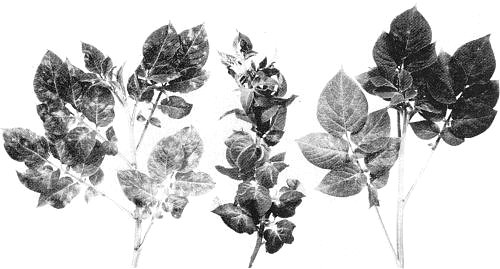
Systemic symptoms in shoots of potato cv. Arran Victory infected with the paracrinkle isolate from King Edward. (Left) primary infection, (centre) secondary infection, (right) healthy.

Local lesions in Datura metel induced by the D 1102 isolate.

Local lesions in Gomphrena globosa induced by the Fortuna isolate.

Local necrotic rings in Nicotiana debneyi induced by the interveinal mosaic isolate.

Virus particles from a leaf dip preparation. Bar represents 500 nm (courtesy of the late Dr J. Brandes).
References list for DPV: Potato virus M (87)
- Bagnall, Larson & Walker, Res. Bull. agric. Exp. Stn Univ. Wis. 198, 45 pp., 1956.
- Bagnall, Wetter & Larson, Phytopathology 49: 435, 1959.
- Bode & Weidemann, Proc. 4th trienn. Conf. Eur. Ass. Potato Res. Brest 1969: 224, 1970.
- Brandes & Wetter, Phytopath. Z. 49: 61, 1963.
- Brandes, Wetter, Bagnall & Larson, Phytopathology 49: 443, 1959.
- Hakkaart, Van Slogteren & De Vos, Tijdschr. PlZiekt. 68: 126, 1962.
- Harrison, Finch, Gibbs, Hollings, Shepherd, Valenta & Wetter, Virology 45: 356, 1971.
- Hiruki, Phytopathology 60: 739, 1970.
- Howard & Wainwright, Nature, Lond. 186: 993, 1960.
- Kahn, Scott, Bozicevich & Vincent, Phytopathology 57: 61, 1967.
- Kassanis, Ann. appl. Biol. 43: 103, 1955.
- Kassanis, J. gen. Microbiol. 15: 620, 1956.
- Kassanis, Ann. appl. Biol. 45: 422, 1957.
- Kassanis, Eur. Potato J. 4: 13, 1961.
- Köhler, Ber. dt. bot. Ges. 66: 63, 1953.
- McKay & Dykstra, Bull. Ore. agric. Exp. Stn 294, 1, 1932.
- Rozendaal & Van Slogteren, Proc. 3rd Conf. Potato Virus Diseases, Lisse-Wageningen 1957: 20, 1958.
- Salaman & Le Pelley, Proc. R. Soc. B 106: 140, 1930.
- Schultz & Folsom, J. agric. Res. 25: 43, 1923.
- Van Slogteren, Proc. 2nd Conf. Potato Virus Diseases, Lisse-Wageningen, 1954: 51, 1955.
- Varma, Gibbs, Woods & Finch, J. gen. Virol. 2: 107, 1968.
- Wetter, Arch. Mikrobiol. 37: 278, 1960.
- Wetter, Z. Naturf. B 22: 1008, 1967.
- Wetter & Brandes, Phytopath. Z. 26: 81, 1956.
- Wetter & Völk, Eur. Potato J. 3: 158, 1960.