Details of DPV and References
DPV NO: 120 July 1973
Family: Virgaviridae
Genus: Tobravirus
Species: Pea early-browning virus | Acronym: PEBV
Pea early-browning virus
B. D. Harrison Scottish Horticultural Research Institute, Invergowrie, Dundee, Scotland
Contents
- Introduction
- Main Diseases
- Geographical Distribution
- Host Range and Symptomatology
- Strains
- Transmission by Vectors
- Transmission through Seed
- Transmission by Grafting
- Transmission by Dodder
- Serology
- Nucleic Acid Hybridization
- Relationships
- Stability in Sap
- Purification
- Properties of Particles
- Particle Structure
- Particle Composition
- Properties of Infective Nucleic Acid
- Molecular Structure
- Genome Properties
- Satellite
- Relations with Cells and Tissues
- Ecology and Control
- Notes
- Acknowledgements
- Figures
- References
Introduction
-
Described by
Bos & Van der Want (1962).
Synonym
- Vroege-verbruiningsvirus van de erwt (Rev. appl. Mycol. 42: 506)
-
An RNA-containing virus with straight tubular particles c. 21 nm wide and of two predominant lengths, c. 105 and 215 nm. Most isolates are readily transmitted by inoculation of sap but others are not. The host range is moderately wide and the vectors are soil-inhabiting nematodes (Trichodorus spp.). Found in western Europe.
Main Diseases
Causes early-browning disease of pea: large necrotic segments develop in the stipules and leaflets (Fig. 1), and necrotic streaks may occur on the stems. Necrosis is usually confined to part of a shoot, or of a leaf, but whole shoots may be killed. Necrotic areas sometimes occur on the pods. The reaction to infection of pea cultivars depends on the cultivar and the virus isolate (Hubbeling & Kooistra, 1963; Harrison, 1966; Van Hoof, 1969). Infected lucerne produces somewhat distorted leaves with chlorotic chevrons (Fig. 2).
Geographical Distribution
Western Europe, especially The Netherlands and England.
Host Range and Symptomatology
At least 30 dicotyledonous species in at least 10 plant families have been infected by inoculation of sap. In many species the virus does not become systemic (Bos & Van der Want, 1962; Gibbs & Harrison, 1964).
-
Diagnostic species
- Chenopodium amaranticolor.
Necrotic lesions 2-5 mm diameter in inoculated leaves (Fig. 5). Some virus isolates tend to produce scattered necrotic lesions in systemically infected leaves but most isolates do not become systemic. - Nicotiana clevelandii. Diffuse chlorotic or necrotic lesions in inoculated
leaves. The first leaves to be invaded systemically may develop necrotic markings
(Fig. 3)
but later-formed leaves are nearly symptomless although infected.
- Phaseolus vulgaris cv. The Prince (French bean). Necrotic lesions up to 3 mm diameter in inoculated primary leaves (but see also under Strains). Rarely infected systemically, developing sporadic necrotic lesions.
- Pisum sativum cv. Onward (pea). Necrotic or chlorotic lesions in inoculated leaves. Systemically infected leaves are stunted and mottled.
- Vicia faba cv. The Sutton (broad bean). British isolates cause diffuse chlorotic and necrotic patches in inoculated leaves. Necrotic streaks form on stems, and tip leaves develop a necrotic wilt. Dutch isolates have not been tested using The Sutton, but cause symptomless systemic infection in cv. Driemaal Wit.
- Phaseolus vulgaris cv. The Prince (French bean). Necrotic lesions up to 3 mm diameter in inoculated primary leaves (but see also under Strains). Rarely infected systemically, developing sporadic necrotic lesions.
-
Propagation species
- Nicotiana clevelandii
is a suitable host for maintaining cultures and as a source of virus for purification.Assay species
- Chenopodium amaranticolor
and Phaseolus vulgaris are the most useful plants for local lesion assay. Cucumis sativus (cucumber; roots only are infected) and pea are useful bait plants for testing transmission by vectors.
Strains
Dutch strain (Bos & Van der Want, 1962). The type strain. Prevalent in the Netherlands; not found in Britain.
British strain (Gibbs & Harrison, 1964). Found in England. Serologically only distantly related to the Dutch type strain. Two variants of the British strain are reported (Harrison, 1966). One of these is transmitted by vectors to the same range of pea cultivars as the type strain; the other infects all cultivars tested. The Dutch apical necrosis isolate of Hubbeling & Hijberts (1968) has some similarities with this second variant.
Italian No. 6 strain (Van Hoof, Maat & Seinhorst, 1966; Cooper & Mayo, 1973). Serologically more closely related to the British strain than is the type strain; produces tiny lesions (resembling those of tobacco rattle virus) in inoculated French bean leaves. Originally obtained in Italy from tobacco bait seedlings in soil containing Trichodorus viruliferus.
Isolates that produce infective RNA but do not produce the characteristic nucleoprotein particles (Gibbs & Harrison, 1964) are found in naturally infected plants: they can also be obtained by using inocula containing only the 215 nm virus particles (Huttinga, 1969). They are poorly transmissible by inoculation of sap but readily transmissible using inocula made with the aid of phenol. Some such isolates produce only tiny lesions in inoculated French bean leaves (Lister, 1967).
Transmission by Vectors
Vectors are stubby root nematodes (Trichodorus spp.). T. teres and T. pachydermus are vectors in The Netherlands (Van Hoof, 1962), and T. anemones, T. primitivus and T. viruliferus in England (Gibbs & Harrison, 1964; Harrison, 1966, 1967). T. anemones and T. similis did not transmit Dutch isolates. Adult male, adult female and juvenile T. primitivus can inoculate the virus, which can be retained for at least 32 days by nematodes kept without plants (Harrison, 1966).
Transmission through Seed
A Dutch isolate is readily seedborne (37%) in pea cv. Rondo. Virus-carrying seeds are wrinkled and discoloured; the virus is easily detected in affected seeds (Bos & Van der Want, 1962). British isolates were transmitted through 1-2% of seed of several pea cultivars (B. D. Harrison, unpublished results).
Transmission by Dodder
No reports.
Serology
Moderately immunogenic. Antisera with precipitin titres of 1/1000 can be obtained. Precipitin tests in mixed liquids are satisfactory.
Relationships
Various degrees of antigenic relatedness are found among different strains. Maat (1963) reported a distant serological relationship between Dutch isolates of pea early-browning and tobacco rattle viruses, and Cooper & Mayo (1973) noted isolates intermediate in other properties between the type strains of these two viruses, which comprise the tobravirus group.
Stability in Sap
In Nicotiana clevelandii sap, the thermal inactivation point (10 min) is 74 to 78°C, dilution end-point about 10-5 and sap remains infective at 20°C for at least a year.
Purification
Collect systemically infected Nicotiana clevelandii leaves about 14 days after inoculation, mince, and store expressed sap for about a month at -15°C. Thaw overnight, heat at 50°C for 10 min, centrifuge at 8000 g for 10 min, and purify and concentrate virus from the supernatant fluid by two cycles of differential centrifugation. Resuspend pellets from high-speed centrifugation in 0.02 M phosphate buffer, pH 7.4 (Cooper & Mayo, 1972).
Properties of Particles
All isolates (except those unable to produce nucleoprotein particles) produce tubular particles of two predominant lengths, usually about 105 nm (S) and 215 nm (L) respectively (Fig. 4). Some Dutch isolates have S particles shorter than 105 nm (Van Hoof, 1969). Infectivity is associated with L particles, but both L and S particles must be inoculated to give progeny particles of both lengths (Harrison, 1966; Huttinga, 1969).
Sedimentation coefficients (s20, w) in svedbergs: 210 (S) and 286 (L).
Buoyant density in CsCl: 1.31 (L and S).
A260/A280: about 1.15.
Particle Structure
Particles are helically constructed. The particle width is about 21.5 nm, core width 4 nm and the pitch of the helix is 2.5 nm (Harrison, 1966; Cooper & Mayo, 1972).
Particle Composition
RNA : Single-stranded, M. Wt 2.5 x 10⁶ (L particles) and 1.3 x 10⁶ (S particles) (Cooper & Mayo, 1972).
Protein: M. Wt of polypeptide subunits, 24,000 (Cooper & Mayo, 1972). The polypeptide in virus preparations stored without bacteriostatic agent becomes converted to a form of apparent M. Wt 21,000, as determined by polyacrylamide gel electrophoresis (Mayo & Cooper, 1973).
Relations with Cells and Tissues
Small groups of particles were observed in the cytoplasm of Nicotiana clevelandii leaf cells (B. D. Harrison & I. M. Roberts, unpublished results).
Notes
Pea early-browning virus occurs mostly on loamy sand soils, which are favoured by its nematode vectors. Unlike tobacco rattle virus, it infects pea systemically. The two viruses differ serologically and have L particles of slightly different length. Tobacco rattle virus differs from most strains of pea early-browning virus by producing only small lesions in inoculated French bean leaves. Other viruses infecting pea can be distinguished by their particle morphology and by the reaction of Chenopodium amaranticolor, Nicotiana clevelandii and Phaseolus vulgaris.
Acknowledgements
Figs 1-3 and 5 from Gibbs & Harrison, 1964; Fig. 4 from Harrison, 1966.
Figures
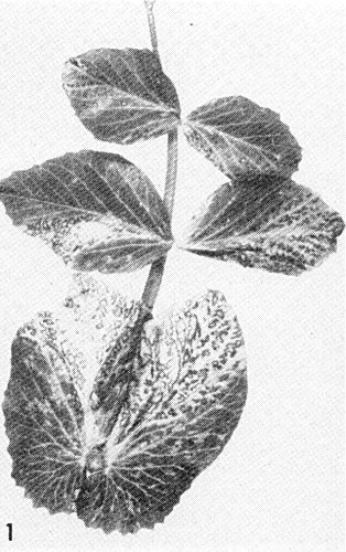
Leaf and stipules of naturally infected pea cv. Emblem.

Leaf of naturally infected lucerne.

Systemically infected Nicotiana clevelandii leaf.

Virus particles in phosphotungstate. Bar represents 100 nm.

Inoculated Chenopodium amaranticolor leaf.
References list for DPV: Pea early-browning virus (120)
- Bos & Van der Want, Tijdschr. PlZiekt. 68: 368, 1962.
- Cooper & Mayo, J. gen. Virol. 16: 285, 1972.
- Cooper & Mayo, Rep. Scott. hort. Res. Inst. 1972: 1973.
- Gibbs & Harrison, Ann. appl. Biol. 54: 1, 1964.
- Harrison, Ann. appl. Biol. 57: 121, 1966.
- Harrison, Rep. Rothamsted Exp. Stn, 1966: 115, 1967.
- Hubbeling & Hijberts, Jversl. Inst. plziektenk. Onderz. 1967: 131, 1968.
- Hubbeling & Kooistra, Zaadbelangen 17: 256, 1963.
- Huttinga, Neth. J. Pl. Path. 75: 338, 1969.
- Lister, Virology 31: 739, 1967.
- Maat, Tijdschr. PlZiekt. 69: 287, 1963.
- Mayo & Cooper, J. gen. Virol. 18: 281, 1973.
- Van Hoof, Tijdschr. PlZiekt. 68: 391, 1962.
- Van Hoof, Meded. Rijksfac. LandbWetensch. Gent. 34: 888, 1969.
- Van Hoof, Maat & Seinhorst, Neth. J. Pl. Path. 72: 253, 1966.