Details of DPV and References
DPV NO: 122 July 1973
Family: Potyviridae
Genus: Potyvirus
Species: Passion fruit woodiness virus | Acronym: PWV
Passionfruit woodiness virus
R. H. Taylor Victorian Plant Research Institute, Burnley, Victoria, Australia
R. S. Greber Department of Primary Industries, Indooroopilly, Queensland, Australia
Contents
- Introduction
- Main Diseases
- Geographical Distribution
- Host Range and Symptomatology
- Strains
- Transmission by Vectors
- Transmission through Seed
- Transmission by Grafting
- Transmission by Dodder
- Serology
- Nucleic Acid Hybridization
- Relationships
- Stability in Sap
- Purification
- Properties of Particles
- Particle Structure
- Particle Composition
- Properties of Infective Nucleic Acid
- Molecular Structure
- Genome Properties
- Satellite
- Relations with Cells and Tissues
- Ecology and Control
- Notes
- Acknowledgements
- Figures
- References
Introduction
- Described by McKnight (1953) and Taylor & Kimble (1964).
- A virus with flexuous particles about 750 x 12 nm, sap-transmissible to a wide range of hosts, particularly Leguminosae, and transmitted by at least two species of aphids in the non-persistent manner.
Main Diseases
Causes mosaic (Fig. 1), ringspots, rugosity and distortion of leaves of Passiflora edulis (passionfruit); the fruits are frequently distorted and the pericarp hard and thick (Fig. 3). The productive life of the plants is greatly decreased. Simmonds (1959) regarded the disease as the most important disease of Passiflora edulis in sub-tropical Australia. However, its importance has declined since the widespread use of vegetatively propagated plants of the tolerant hybrid, P. edulis x P. edulis f. flavicarpa. The virus also occurs naturally in some tropical legumes, including Arachis hypogea, Centrosema pubescens, Crotalaria usaramoensis, Glycine max and Phaseolus atropurpureus, but is of minor importance in these species.
Geographical Distribution
Reported from the Australian States of Queensland, New South Wales and Western Australia, and from Surinam (J. J. de Wijs, pers. comm.).
Host Range and Symptomatology
Reported to infect 44 dicotyledonous plant species in five families (Taylor & Kimble, 1964; Teakle & Wildermuth, 1967). Hosts include 10 species of Passiflora and 18 in the Leguminosae.
- Diagnostic species
- Passiflora edulis
(passionfruit). Mosaic (Fig. 1), rugosity and distortion, with yellow spots on older leaves. The woody fruit symptom (Fig. 3) may also be caused by cucumber mosaic virus or by insect feeding injury and is of limited diagnostic value. - Passiflora edulis f. flavicarpa (golden passionfruit) and
P. edulis x P. edulis f. flavicarpa (hybrid passionfruit).
Pale green to chlorotic ringspots occur on spring growth together with leaf
mosaic (Fig. 2).
- Phaseolus lathyroides. Inoculated leaves show chlorotic to necrotic local lesions, followed by systemic infection with leaf epinasty, vein necrosis, leaf abscission and terminal necrosis.
- Phaseolus vulgaris (French bean). Depending on the cultivar, symptoms in inoculated leaves range from faint local lesions to veinal necrosis (Fig. 5). In cv. Bountiful the local lesions are chlorotic with normal strains and brown necrotic with severe strains. The first systemically infected leaves show a faint mottle which often gives way to a severe blister at a later stage of infection. In some cultivars necrosis and mottling of the pods occurs.
- Phaseolus lathyroides. Inoculated leaves show chlorotic to necrotic local lesions, followed by systemic infection with leaf epinasty, vein necrosis, leaf abscission and terminal necrosis.
- Propagation species
- Passiflora edulis
may be used to maintain all except the tip blight strain which may be maintained in the more tolerant host Passiflora suberosa. Phaseolus vulgaris cv. Bountiful is a good source of virus for purification.- Assay species
- Small countable, rusty-red ringspots are produced on inoculated primary leaves of Dolichos biflorus. Chlorotic lesions which may be counted with difficulty are produced in Phaseolus vulgaris cv. Bountiful. Very small seedlings of Passiflora edulis or Phaseolus lathyroides are useful for testing transmission by vectors.
Strains
Several strains can be distinguished by severity of symptoms in Passiflora edulis. Greber (1966) described a strain which is mild in Passiflora suberosa but causes tip blight and complete loss of crop in Passiflora edulis. Greber (1971) described a strain which occurs naturally in Centrosema pubescens and causes mild mosaic disease in Passiflora edulis. Mild strains effectively cross-protect against virulent strains in Passiflora edulis.
Transmission by Vectors
Transmitted in the non-persistent manner by the aphids Myzus persicae (Taylor, 1959) and Aphis gossypii (Greber, 1966). No other species have been tested as vectors.
Transmission through Seed
Observations on Passiflora seedlings grown for experimental or commercial purposes give no evidence of seed transmission.
Serology
Strongly immunogenic. Antisera with titres up to 1/1024 were obtained by Taylor & Kimble (1964) by subcutaneous injections of virus in Freund’s adjuvant at several sites followed ten days later by an intravenous injection. Serological precipitates in mixed liquids occur in a few minutes and are of the flocculent type.
Relationships
All strains tested appear to be closely related serologically. The virus is similar to bean yellow mosaic virus in shape, physical properties and symptoms in Phaseolus vulgaris, but it does not appear to be serologically related to this virus or to the pea mosaic strain of it (Taylor & Kimble, 1964; Teakle & Wildermuth, 1967). Its biological and physical properties place it in the potyvirus group.
Stability in Sap
In Phaseolus vulgaris sap the thermal inactivation point (10 min) is 55-60°C, dilution end-point 10-4 to 10-5 and inactivation in vitro occurs in 3-4 days at 18°C.
Purification
The virus is stable and is readily purified from inoculated primary leaves of Phaseolus vulgaris cv. Bountiful by the following method. Blend each 100 g of infected leaf tissue in 100 ml phosphate buffer (0.3 M, pH 7.6) containing 0.1% thioglycollic acid. Squeeze out the fluid through cheesecloth, add n-butanol to 8.5% (v/v), place extract in an ice-bath for 1-2 hr, centrifuge at low speed and concentrate by ultracentrifugation. To minimize aggregation, resuspend pellets in borate buffer (0.05 M, pH 8.2) and not in phosphate buffer. Density gradient centrifugation may be used to purify the virus further. Yields are high compared with most other potyviruses. Preparations showing strong birefringence may be obtained from 200 g leaf.
Properties of Particles
No information.
Particle Structure
Particles are flexuous filaments about 12 nm wide (Fig. 6). In partially purified preparations Taylor & Kimble (1964) found a normal length of 670 nm, whereas Teakle & Wildermuth (1967) reported a figure of 745 nm. More recently R. S. Greber (unpublished) measured the lengths of 100 particles in Phaseolus vulgaris sap, using catalase crystals and tobacco mosaic virus as standards, and obtained a figure of 700 nm.
Particle Composition
Not known.
Relations with Cells and Tissues
Pinwheel inclusions and particles occur frequently in the mesophyll and vascular parenchyma of Phaseolus vulgaris (Fig. 4).
Notes
A woodiness disease of passionfruit was described by Cobb (1901) but as both passionfruit woodiness and cucumber mosaic viruses may cause woody fruit and leaf mosaic (Taylor, 1959), the authenticity of records prior to that of McKnight (1953) is in doubt. However, passionfruit woodiness virus was rare in P. edulis in Queensland in 1927 and widespread in 1932 (Simmonds, 1959) suggesting that it was introduced into that State about 1927. Its absence from cool, temperate areas of Australia and the rare occurrence of cucumber mosaic virus in Queensland suggest that ecological factors influence the distribution of both viruses. Experimentally, a mixed infection by the two viruses in P. edulis causes a very severe disease (Taylor, 1959), but naturally occurring mixed infections are not always so virulent.
To differentiate the two viruses it is best to use the electron microscope; the filamentous particles of passionfruit woodiness virus are usually readily found in the sap of infected plants. The diagnosis may then be confirmed by transmissions to diagnostic hosts. Negative results suggest the presence of cucumber mosaic virus which, except in mixed infection with passionfruit woodiness virus, exists in very low concentration in seedlings of Passiflora edulis and is extremely difficult to transmit to diagnostic hosts by aphid or sap inoculation. However, when Passiflora edulis scions infected with cucumber mosaic virus are grafted onto Passiflora caerulea stock the scions develop a severe necrotic disease and often ultimately die. The virus reaches a high enough concentration in both stock and scion for transfers to be made from either to diagnostic hosts.
Passionfruit woodiness virus could be confused with passionfruit latent virus (Schnepf & Brandes, 1962), which has slightly flexuous particles about 650 nm long, unless care is taken to check particle shape and length. Mixed infections of the two viruses have not been reported.
A virus reported in passionfruit in Nigeria (Martini, 1962) may be related but serological evidence is not available.
The use of mild strains to protect plants from more virulent ones in field conditions was suggested by McKnight (1953) and applied by Simmonds (1959); it appears to be one of the few instances of this method of control having been economically useful. More recently, partial control has been obtained by the use of the hybrid passionfruit which, like its Passiflora edulis f. flavicarpa parent, produces distorted fruit only under adverse conditions or when infected with an unusually virulent strain of the virus.
Figures
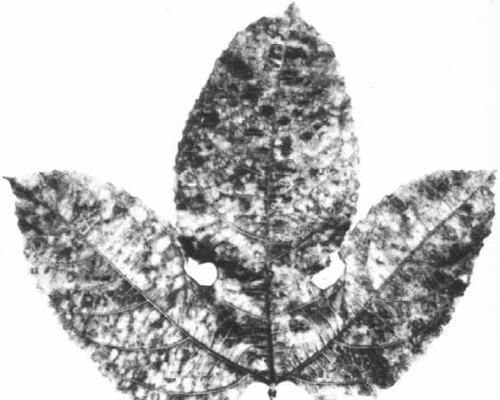
Mosaic in Passiflora edulis.

Ringspots and mosaic in Passiflora edulis x P. edulis f. flavicarpa.

Woody fruit of Passiflora edulis.

Pinwheel inclusions and bundles in mesophyll cells of Phaseolus vulgaris cv. Bountiful. Bar represents 200 nm.

Inoculated leaf of Phaseolus vulgaris cv. Sutter’s Pink showing necrotic local lesions and spreading veinal necrosis.

Virus particles from sap of Phaseolus vulgaris cv. Bountiful. Bar represents 200 nm.
References list for DPV: Passionfruit woodiness virus (122)
- Cobb, Agric. Gaz. N. S. W. 12: 407, 1901.
- Greber, Qd. J. agric. Anim. Sci. 23: 533, 1966.
- Greber, Qd. J. agric. Anim. Sci. 28: 115, 1971.
- Martini, Ann. appl. Biol. 50: 163, 1962.
- McKnight, Qd. J. agric. Sci. 10: 4, 1953.
- Schnepf & Brandes, Phytopath. Z. 43: 102, 1962.
- Simmonds, Qd. J. agric. Sci. 16: 371, 1959.
- Taylor, J. Aust. Inst. agric. Sci. 25: 71, 1959.
- Taylor & Kimble, Aust. J. agric. Res. 15: 560, 1964.
- Teakle & Wildermuth, Qd. J. agric. Anim. Sci. 24: 173, 1967.