Details of DPV and References
DPV NO: 136 July 1974
Family: Closteroviridae
Genus: Closterovirus
Species: Carnation necrotic fleck virus | Acronym: CNFV
Carnation necrotic fleck virus
T. Inouye College of Agriculture, University of Osaka Prefecture, Sakai, Osaka, Japan
Contents
- Introduction
- Main Diseases
- Geographical Distribution
- Host Range and Symptomatology
- Strains
- Transmission by Vectors
- Transmission through Seed
- Transmission by Grafting
- Transmission by Dodder
- Serology
- Nucleic Acid Hybridization
- Relationships
- Stability in Sap
- Purification
- Properties of Particles
- Particle Structure
- Particle Composition
- Properties of Infective Nucleic Acid
- Molecular Structure
- Genome Properties
- Satellite
- Relations with Cells and Tissues
- Ecology and Control
- Notes
- Acknowledgements
- Figures
- References
Introduction
-
Described by
Inouye & Mitsuhata (1973).
A virus with very flexuous elongated particles c. 1400-1500 nm long. Transmitted by aphids in a semi-persistent manner but with difficulty by inoculation of sap. It seems to infect only species of Caryophyllaceae. Found in Japan.
Main Diseases
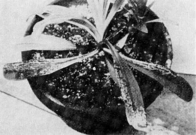
Causes greyish-white or reddish-purple necrotic flecks, streaks or spots in carnations (Fig. 1). Often found in mixed infection with other carnation viruses. The symptoms are often masked at lower temperatures.
Geographical Distribution
Japan (Inouye & Mitsuhata, 1973).
Host Range and Symptomatology
Only hosts known are 3 species in Caryophyllaceae. Twelve species in Chenopodiaceae, Compositae, Cruciferae, Cucurbitaceae, Leguminosae and Solanaceae were not infected when inoculated by aphids and/or with plant sap (Inouye & Mitsuhata, 1973).
-
Diagnostic species
- Dianthus caryophyllus
(carnation). Greyish-white necrotic spots and flecks, 2-3 weeks after inoculation by aphids (Fig. 1), sometimes followed by reddish-purple discoloration of leaves. Severely affected seedlings are stunted and killed, but most chronically infected plants are symptomless. - Dianthus barbatus. Veinal chlorosis and necrosis appears on fully expanded young leaves 2-3 weeks after inoculation by aphids, often appearing as yellow net symptoms (Fig. 3). Affected leaves eventually show reddish discoloration and tip necrosis (Fig. 2). Leaves produced subsequently show symptoms only at the leaf tips, and leaves developing still later are almost symptomless. Mechanically inoculated plants develop necrotic lesions in inoculated leaves (Fig. 4), but only a few become infected systemically, producing faint veinal chlorosis in the upper leaves (Inouye & Mitsuhata, 1973).
-
Propagation species
- Dianthus barbatus
is a suitable host for maintaining cultures.Assay species
- Dianthus barbatus
gives local lesions following sap inoculation; it is also good for tests by aphid inoculation.
Strains
No strains reported.
Transmission by Vectors
Transmitted by the aphid Myzus persicae (Inouye & Mitsuhata, 1973) in a semi-persistent manner. Probability of transmission increases with increase of acquisition feed beyond 4 h and of test feed beyond 30 min. Vector aphids can retain the virus for up to 2 days.
Transmission through Seed
Not tested.
Transmission by Dodder
Not tested.
Serology
No antisera have been prepared.
Relationships
In having very flexuous particles over 1 µm long and in its transmission by aphids in the semi-persistent manner the virus resembles beet yellows (Russell, 1970), citrus tristeza (Price, 1970), festuca necrosis (Schmidt et al., 1963), and wheat yellow leaf (Inouye et al., 1973) viruses. The virus also resembles beet yellows, citrus tristeza and wheat yellow leaf viruses in its relations with tissues.
Stability in Sap
In Dianthus barbatus sap, the thermal inactivation point is between 40 and 45°C, the dilution end-point is around 10-4, and longevity in vitro is between 2 and 4 days at 20°C (T. lnouye, unpublished data).
Purification
Flexuous rods have been partially purified using the following method (T. Inouye. unpublished data). Express juice from frozen leaves of infected D. barbatus, with mortar and pestle, in 0.1 M phosphate buffer pH 7.6 containing 0.1% thioglycollic acid. Clarify juice using ether, then carbon tetrachloride. Precipitate the virus adding polyethylene glycol M. Wt 6000 to 4% (w/v) and NaCl to 0.3 M. Centrifuge at low speed and resuspend pellets in 0.01 M phosphate buffer pH 7.6. Concentrate by differential centrifugation, resuspending the pellets from high speed centrifugation in 0.01 M phosphate buffer.
Properties of Particles
Unknown.
Particle Structure
Particles are flexuous filaments c. 1400-1500 nm long and 12-13 nm in diameter, and are helically constructed with the pitch of the basic helix c. 3.4 nm (Fig. 6) (Inouye & Mitsuhata, 1973).
Particle Composition
Unknown.
Relations with Cells and Tissues
Causes necrosis of some phloem cells. Masses of virus particles and/or vesicular structures are observed in some phloem and epidermal cells (Fig. 7) (Inouye & Mitsuhata, 1973). Cellular inclusions (X-bodies) are visible by light microscopy in epidermal strips of infected D. barbatus stained with phloxine-methylene blue (Fig. 5) (Inouye & Mitsuhata, 1973).
Notes
Carnation necrotic fleck virus can be distinguished from two other carnation viruses having elongated particles, carnation latent (Wetter, 1971) and carnation vein mottle (Hollings & Stone, 1971), by its longer and more flexuous particles, by its semi-persistent mode of transmission by aphids, and by producing necrotic local lesions following sap inoculation to D. barbatus. In this host, carnation latent virus causes no symptoms and carnation vein mottle virus induces conspicuous leaf mottling. Cross bands observable in particles of carnation necrotic fleck virus negatively stained with phosphotungstate may also be useful for distinguishing this virus from other elongated virus particles occurring in carnation.
Figures

Systemic necrotic flecks and spots in carnation.
Systemic veinal necrosis and leaf discoloration in Dianthus barbatus.
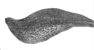
A leaf of D. barbatus, showing veinal chlorosis followed by veinal necrosis.
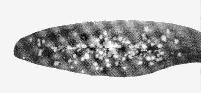
Local lesions in D. barbatus induced by sap inoculation.
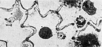
Cellular inclusions (X-bodies) in epidermal strip of D. barbatus.

Parts of virus particles negatively stained with phosphotungstate; bar represents 100 nm.

Ultrathin section of a phloem cell of infected D. barbatus, showing aggregates of virus particles and vesicular structures; bar represents 1 µm.
References list for DPV: Carnation necrotic fleck virus (136)
- Hollings & Stone, CMI/AAB Descriptions of Plant Viruses 78, 4 pp., 1971.
- Inouye & Mitsuhata, Ber. Ohara Inst. landw. Biol. 15: 195, 1973.
- Inouye, Mitsuhata, Heta & Hiura, Nogaku Kenkyu 55: 1, 1973.
- Price, CMI/AAB Descriptions of Plant Viruses 33, 3 pp., 1970.
- Russell, CMI/AAB Descriptions of Plant Viruses 13, 3 pp., 1970.
- Schmidt, Richter, Hertzsch & Klinkowski. Phytopath. Z. 47: 66, 1963.
- Wetter, CMI/AAB Descriptions of Plant Viruses 61, 4 pp., 1971.