Details of DPV and References
DPV NO: 142 October 1975
Family: Secoviridae
Genus: Nepovirus
Species: Mulberry ringspot virus | Acronym: MRSV
Mulberry ringspot virus
T. Tsuchizaki Institute for Plant Virus Research, Chiba, Japan
Contents
- Introduction
- Main Diseases
- Geographical Distribution
- Host Range and Symptomatology
- Strains
- Transmission by Vectors
- Transmission through Seed
- Transmission by Grafting
- Transmission by Dodder
- Serology
- Nucleic Acid Hybridization
- Relationships
- Stability in Sap
- Purification
- Properties of Particles
- Particle Structure
- Particle Composition
- Properties of Infective Nucleic Acid
- Molecular Structure
- Genome Properties
- Satellite
- Relations with Cells and Tissues
- Ecology and Control
- Notes
- Acknowledgements
- Figures
- References
Introduction
- Described by Tsuchizaki, Hibino & Saito (1971).
- An RNA-containing virus with isometric particles 22-25 nm in diameter spreading naturally in Japan. It is readily sap-transmissible, has a moderate host range and is transmitted by the nematode, Longidorus martini.
Main Diseases
Found naturally only in mulberry, in which it usually causes mosaic and ringspot symptoms (Fig.1). Some isolates are associated with leaf enations.
Geographical Distribution
Japan.
Host Range and Symptomatology
Restricted in nature to mulberry. In limited host range studies, it infected 10 species in 5 dicotyledonous families.
- Diagnostic species
- Morus alba (mulberry). Systemically infected leaves develop mosaic,
ringspots (Fig. 1) or enations
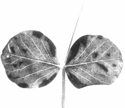
- Vigna sinensis cv. Black Eye (cowpea). Necrotic spots or ringspots in inoculated leaves (Fig. 2), followed by mosaic or necrosis of leaves and necrosis of stems.
- Glycine max (soybean). Systemically infected leaves develop mosaic or chlorotic spots.
- Pisum sativum (pea). Chlorotic spotting or mosaic of systemically infected leaves (Fig. 3).
- Chenopodium quinoa. Indistinct chlorotic local lesions may form; systemically infected leaves show mosaic.
- Vigna sinensis cv. Black Eye (cowpea). Necrotic spots or ringspots in inoculated leaves (Fig. 2), followed by mosaic or necrosis of leaves and necrosis of stems.
- Propagation species
- Vigna sinensis cv. Black Eye is a suitable host for maintaining cultures.
- Assay species
- Vigna sinensis cv. Black Eye, although not always satisfactory, can be used for local lesion assays.
Strains
No strains reported.
Transmission by Vectors
Transmitted by the nematode, Longidorus martini, but not by a species of Xiphinema (Yagita & Komuro, 1972). Infested soil containing L. martini was still infective after storage, in the absence of plants, for 14-17 months at room temperature or for more than 30 months at 0-9°C (H. Yagita, unpublished).
Transmission through Seed
About 10% of progeny seedlings of Glycine max cv. Mikawashima are infected (Tsuchizaki et al., 1971).
Transmission by Dodder
Not reported.
Serology
Moderately antigenic, giving antisera with titres up to 1/320. In gel-diffusion tests it reacts with antisera to produce a distinct single band of precipitate.
Relationships
Mulberry ringspot virus did not react with antisera to arabis mosaic, cherry leaf roll (type and golden elderberry strains), grapevine fanleaf, grapevine chrome mosaic, raspberry ringspot, strawberry latent ringspot, tobacco ringspot, tomato black ring or tomato ringspot viruses (B. D. Harrison, unpublished results).
Stability in Sap
In sap of Glycine max, the virus lost infectivity after 10 min at 50-60°C, storage at room temperature for 3-5 days, or dilution to 10-3-10-4 (Tsuchizaki et al., 1971).
Purification
Harvest cowpea plants about 2 weeks after inoculation, then homogenize at 4°C in two volumes of 0.5 M citrate buffer (pH 7.0) containing 0.1% thioglycollic acid. Express juice through cheesecloth, and add 20 ml carbon tetrachloride to every 100 ml extract. Shake the extract for 15 min, and clarify by low-speed centrifugation. Concentrate the virus by three cycles of differential centrifugation. Resuspend the pellets from high speed centrifugation in 0.01 M citrate buffer. Purify further by sucrose density-gradient centrifugation (Tsuchizaki et al., 1971).
Properties of Particles
The particles are all the same size but sediment as three components, apparently empty protein shells (T) and two kinds of nucleoprotein (M and B) (Tsuchizaki et al., 1971).
Sedimentation coeflicients (s20,w) (svedbergs): 50 (T), 96 (M), 126 (B) (T. Tsuchizaki, unpublished data).
Other physical properties are unknown.
Particle Structure
Particles are isometric, 22-25 nm in diameter. Electron micrographs show some particles completely, some partially and some not penetrated by negative stain (Fig. 4).
Particle Composition
No quantitative information, but the particles contain RNA (T. Tsuchizaki, unpublished data).
Relations with Cells and Tissues
In infected cowpea and mulberry leaves, electron microscopy revealed tubules containing a single row of spherical virus-like particles (Fig. 5), and characteristic vesicular inclusion bodies (Tsuchizaki et al., 1971).
Notes
Similarity of properties and transmissibility by a nematode place mulberry ringspot virus in the nepovirus group, although its particles are perhaps smaller than those of other viruses in the group. Symptoms in experimental hosts may be confused with those caused by other nepoviruses. Identification should be made by serology. An elongated virus is frequently found in mulberries naturally infected with this virus. The two viruses differ in their infectivities for cowpea or soybean.
Figures
References list for DPV: Mulberry ringspot virus (142)
- Tsuchizaki, Hibino & Saito, Ann. phytopath. Soc. Japan 37: 266, 1971.
- Yagita & Komuro, Ann. phytopath. Soc. Japan 38: 275, 1972.


