Details of DPV and References
DPV NO: 143 October 1975
Family: Potyviridae
Genus: Bymovirus
Species: Barley yellow mosaic virus | Acronym: BaYMV
There is a more recent description of this virus: DPV 374
Barley yellow mosaic virus
T. Inouye College of Agriculture, University of Osaka Prefecture, Sakai, Osaka, Japan
Y. Saito Institute for Plant Virus Research, Ministry of Agriculture, Aoba Cho, Chiba, Japan
Contents
- Introduction
- Main Diseases
- Geographical Distribution
- Host Range and Symptomatology
- Strains
- Transmission by Vectors
- Transmission through Seed
- Transmission by Grafting
- Transmission by Dodder
- Serology
- Nucleic Acid Hybridization
- Relationships
- Stability in Sap
- Purification
- Properties of Particles
- Particle Structure
- Particle Composition
- Properties of Infective Nucleic Acid
- Molecular Structure
- Genome Properties
- Satellite
- Relations with Cells and Tissues
- Ecology and Control
- Notes
- Acknowledgements
- Figures
- References
Introduction
-
Described by Ikata & Kawai (1940).
A virus with slightly flexuous filamentous particles of two lengths, c. 275 and 550 nm, and both 13 nm wide. It is sap-transmissible and soil-borne; the fungus Polymyxa graminis is thought to be a vector. The only host known is barley. Found in Japan.
Main Diseases
Causes mosaic in barley (Fig. 1); sometimes the leaves show complete yellowing with necrotic patches; plants are stunted. The severity of symptoms depends on the cultivar of barley and the environmental conditions. Symptoms are usually not produced above 18°C. They usually appear in early spring but tend to disappear as the weather becomes warmer.
Geographical Distribution
Japan.
Host Range and Symptomatology
The only host known is Hordeum vulgare (barley). Transmitted through infective soil, and by inoculation of sap. The species of plants tested but not infected include Triticum aestivum (wheat), Avena sativa (oat), Oryza sativa (rice), Chenopodium amaranticolor and Nicotiana tabacum (tobacco).
-
Diagnostic species
- Hordeum vulgare
(barley). The virus is transmissible to barley, though not readily, by inoculation with plant sap and keeping test plants below about 18°C. Increased infectivity was observed when inoculum was made by grinding diseased leaves in 10-3 M KCN, Na-DIECA, or Na-azide (Takanashi et al., 1967), 3% K2HPO4 (Takahashi et al., 1968), or 10-4 M p-nitrophenol (Kusaba et al., 1971).Propagation species
- Hordeum vulgare.
Assay species
- No local lesion host is known. The virus may be assayed by finding the percentage of Hordeum vulgare plants infected using different concentrations of inoculum.
Strains
No special strains have been described, but differences in varietal resistance of barley to different virus isolates (Takahashi et al.,1968) and in different infective soils (Saito & Okamoto, 1964; Kusaba et al.,1971) have been observed.
Transmission by Vectors
Kusaba et al. (1971)
suggested that a fungus, Polymyxa graminis, is the vector of
the virus because (1) steamed soil became infective on addition of
resting spores of the fungus
collected from roots of barley naturally infected with the virus,
(2) virus transmission was not
prevented by treating resting spores of the fungus with 10% Teepol,
5% trisodium orthophosphate, or
at pH 2.3, (3) virus-free fungus acquired the ability to transmit when it was grown in plants
infected with virus by inoculation of sap,
(4) the infectivity of suspensions prepared from naturally
infected roots was closely related to the number of zoospores of the fungus that they contained.
Infectivity in air-dried soil was retained for over 5 years (Yasu & Yoshino, 1964).
Transmission through Seed
Not found (Yasu & Yoshino, 1964).
Transmission by Dodder
Not reported.
Serology
Serum from a rabbit injected intravenously, then intramuscularly, with partially purified virus reacted with the virus to a titre of 1/1280 in complement fixation tests (Usugi & Saito, 1970).
Relationships
In complement fixation tests, barley yellow mosaic virus is serologically related to wheat yellow mosaic and rice necrosis mosaic viruses (Usugi & Saito, 1970). It resembles rice necrosis mosaic (Inouye, 1968, 1970), wheat yellow mosaic (Saito et al., 1966) and wheat spindle streak mosaic (Slykhuis, 1970; Hooper & Wiese, 1972) viruses, in its transmissibility through soil, its particle morphology, and in inducing pinwheel-type inclusions and characteristic membranous network structures in the cytoplasm of infected plant cells.
Stability in Sap
The dilution end-point is less than 10-2 (Kusaba et al.,1971; Yasu & Yoshino, 1964). The thermal inactivation point and longevity in vitro are not known. Infectivity of dried leaf tissue is retained for two years at 5-10°C (Miyamoto, 1958).
Purification
Usugi & Saito (1970): homogenize infected leaf tissue in 0.1 M citrate buffer (pH 7.0), clarify by adding carbon tetrachloride, and concentrate by two or three cycles of differential centrifugation. Two virus-containing bands form in sucrose density gradient centrifugation. Highly purified virus preparations were obtained by equilibrium centrifugation in CsCl, only one band being formed (Usugi & Saito, 1975).
Properties of Particles
The ultraviolet absorption spectrum is typical for nucleoprotein, having a maximum at 265-270 nm with a hump at 290 nm.
A260/A280: 1.14.
Buoyant density in CsCl: 1.29 g/ml (Usugi & Saito, 1975).
Particle Structure
Particles (Fig. 2) of barley yellow mosaic virus are slightly flexuous filaments 13 nm in diameter and having two modal lengths of 275 and 550 nm; both lengths are found in leaf dips and in partially purified virus preparations (Inouye, 1964, 1968).
Particle Composition
Unknown.
Relations with Cells and Tissues
X-bodies can be found in cells of epidermal strips of infected leaves (Fig. 3). In ultrathin sections, prominent membranous network structures, pinwheel-type inclusions, and loosely banded aggregates of virus particles are often observed in the cytoplasm (Saito et al.,1966) (Fig. 4).
Notes
Barley yellow mosaic virus can be distinguished from other viruses with elongated particles that infect barley in Japan by particle morphology and infectivity for Chenopodium amaranticolor or C. quinoa. Barley stripe mosaic (Atabekov & Novikov, 1971) and soil-borne wheat mosaic (Brakke, 1971) viruses have straight rod-shaped particles and cause local lesions in the inoculated leaves of the Chenopodium spp. whereas barley yellow mosaic virus has more slender particles and does not infect these hosts.
Figures

Barley leaves; (left) two leaves showing mottle with necrotic patches; (centre) two leaves showing mottle; (right) healthy leaf.

Virus particles from a partially purified preparation shadowed with chromium. Bar represents 1 µm.

X-bodies in epidermal cells of barley. (N) nucleus; (X) X-body.
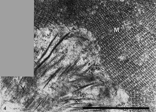
Section of infected leaf cell. (V) loosely banded aggregates of virus particles; (M) membranous network structure. Bar represents 1 µm.
References list for DPV: Barley yellow mosaic virus (143)
- Atabekov & Novikov, CMI/AAB Descriptions of Plant Viruses 68, 4 pp., 1971.
- Brakke, CMI/AAB Descriptions of Plant Viruses 77, 4 pp., 1971.
- Hooper & Wiese, Virology 47: 644,1972.
- Ikata & Kawai, Noji Kairyo Shiryo 154, 123 pp., 1940.
- Inouye, Nogaku Kenkyu 50: 117, 1964.
- Inouye, Ann. phytopath. Soc. Japan 34: 301, 1968.
- Inouye, Ann. phytopath. Soc. Japan 36: 186, 1970.
- Kusaba, Toyama, Yumoto & Tatebe, Spec. Bull. Tottori agric. exp. Stn 2, 208 pp., 1971.
- Miyamoto, Ann. phytopath. Soc. Japan 23: 199, 1958.
- Saito & Okamoto, Bull. natn. Inst. agric. Sci., Tokyo C 17: 75, 1964.
- Saito, Ueda & Inaba, Ann. phytopath. Soc. Japan 32: 87, 1966.
- Slykhuis, Phytopathology 60: 319, 1970.
- Takahashi, Inouye, Hayashi, Moriya, Hirao & Mitsuhata, Nogaku Kenkyu 52: 65, 1968.
- Takanashi, Saito & Iwata, Ann. phytopath. Soc. Japan 33: 43, 1967.
- Usugi & Saito, Ann. phytopath. Soc. Japan 36: 375, 1970.
- Usugi & Saito, Ann. phytopath. Soc. Japan 41: 87, 1975.
- Yasu & Yoshino, Bull. Saitama agric. exp. Stn 24: 1, 1964.