Details of DPV and References
DPV NO: 168 September 1976
Family: Tymoviridae
Genus: Tymovirus
Species: Desmodium yellow mottle virus | Acronym: DYMoV
Desmodium yellow mottle virus
H. A. Scott Department of Plant Pathology, University of Arkansas, Fayetteville, Arkansas 72701, USA
Contents
- Introduction
- Main Diseases
- Geographical Distribution
- Host Range and Symptomatology
- Strains
- Transmission by Vectors
- Transmission through Seed
- Transmission by Grafting
- Transmission by Dodder
- Serology
- Nucleic Acid Hybridization
- Relationships
- Stability in Sap
- Purification
- Properties of Particles
- Particle Structure
- Particle Composition
- Properties of Infective Nucleic Acid
- Molecular Structure
- Genome Properties
- Satellite
- Relations with Cells and Tissues
- Ecology and Control
- Notes
- Acknowledgements
- Figures
- References
Introduction
-
Described by
Walters & Scott (1972).
An RNA-containing virus with isometric particles c. 30 nm in diameter which sediment as two components in the ultracentrifuge. It is restricted to legumes and is readily sap-transmissible. No vector known. Found in USA.
Main Diseases
Causes yellow mottling and some leaf deformity in Desmodium spp. (tick trefoil). The virus has not been obtained from plants of economic importance.
Geographical Distribution
Arkansas, USA.
Host Range and Symptomatology
Only hosts known are members of the Leguminosae. Transmitted readily by sap inoculation.
-
Diagnostic species
- Phaseolus vulgaris
(bean) cv. Great Northern and Vigna sinensis (cowpea) cv. Monarch. Very mild systemic chlorotic mottling. - P. vulgaris cv. Black Valentine, Bountiful and Pinto. Occasional local lesions on inoculated leaves.
-
Propagation species
- Phaseolus vulgaris
cv. Great Northern.Assay species
- Desmodium tortuosum
gives satisfactory local lesions (Fig. 1).
Strains
None reported.
Transmission by Vectors
No vector reported. The bean leaf beetle, Cerotoma trifurcata, the spotted cucumber beetle, Diabrotica undecimpunctata, a weevil, Apion roseae, and the green peach aphid, Myzus persicae, did not transmit the virus (Walters & Scott, 1972).
Transmission through Seed
No report.
Transmission by Dodder
No report.
Serology
The virus is strongly immunogenic. A single band is formed in gel diffusion tests (Fig. 4). Good reactions are obtained with crude sap.
Relationships
The virus belongs to the tymovirus group and is serologically distantly related to turnip yellow mosaic (Scott & Moore, 1972), andean potato latent, dulcamara mottle, ononis yellow mosaic, scrophularia mottle and wild cucumber mosaic viruses (Koenig & Givord, 1974; Koenig, 1976). The virus is closely related to kennedya yellow mottle, okra mosaic, cocoa yellow mosaic and clitoria yellow vein viruses (Koenig, 1976).
Stability in Sap
In Great Northern bean sap, the virus is infective after 10 min at 70°C, but not at 75°C, after dilution to 10-7 but not to 10-8 and after 38 days but not 44 days at 20°C.
Purification
The virus is easily purified by one of the following methods:
1. A modification of Steere’s butanol-chloroform method (Walters & Scott, 1972).
2. Extract tissue with 0.2 M NaH2PO4 (1 gm/2 ml) and squeeze through cheesecloth. Adjust extract to pH 5.0 with 0.1 N HCl and centrifuge at low speed. Subject supernatant fluid to 3 cycles of alternate high and low speed centrifugation, resuspending high speed pellets in 0.01 M phosphate buffer, pH 7.2 (Scott & Moore, 1972).
3. The bentonite method as described by Dunn & Hitchborn (1965) (Koenig & Givord, 1974).
Properties of Particles
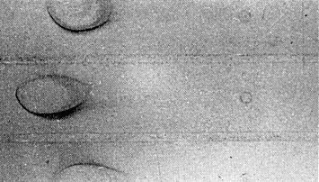
Sedimentation behaviour typical of members of the tymovirus group, i.e. two components (Fig. 2), top (empty protein shells) 54 S and bottom (nucleoprotein) 114 S. Calculation not made at infinite dilution.
In immunoelectrophoresis, using Tris-sodium barbital buffer, pH 8.6, the virus migrated towards the anode as a single component (Fig. 5).
Particle Structure
Particles are isometric c. 30 nm in diameter (Fig. 3) with icosahedral symmetry. Top component particles are penetrated by 2% phosphotungstate, pH 6.8.
Particle Composition
Nucleic acid: RNA, single-stranded, 35% of the particle weight (calculated according to Reichmann, 1965). One centrifugal component, 23.3 S, in 0.01 M phosphate-0.1 M KCl, pH 7.0; 17.7 S when heated in 1% formaldehyde. M. Wt c. 2 x 10⁶. Molar percentages of nucleotides: G16.5, A22.5, C37.2, U23.8 (Scott & Moore, 1972).
Protein: No report.
Relations with Cells and Tissues
Crystals of virus-like particles occur in the nucleus, cytoplasm and often in the vacuoles of cells of infected Phaseolus vulgaris cv. Favorit (Fig. 6) (D. Lesemann, personal communication). Peripheral vesicles bounded by a double membrane occur in the chloroplasts, as shown for other tymoviruses.
Notes
Of the four adequately described viruses isolated from naturally-infected Desmodium spp., desmodium yellow mottle virus may be distinguished by particle morphology from desmodium mosaic virus which was identified in Florida, USA, as a flexuous filamentous virus belonging to the potyvirus group (Edwardson et al., 1970). It is readily distinguished from bean pod mottle (Moore, Scott & Walters, 1969) and cowpea chlorotic mottle (Walters & Dodd, 1969) viruses found in Arkansas, USA, by serology.
Figures
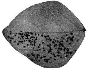
Local lesions on Desmodium tortuosum.

Schlieren patterns obtained by analytical ultracentrifugation comparing purified turnip yellow mosaic (upper) and desmodium yellow mottle (lower) viruses.

Virus particles stained with phosphotungstate. Bar represents 100 nm.

Reciprocal gel diffusion tests comparing turnip yellow mosaic virus (antiserum in centre well at left) and desmodium yellow mottle virus (antiserum in centre well at right). Homologous antigens in top and alternate wells.
Immunoelectrophoretic comparison of turnip yellow mosaic virus (top and bottom bands) with desmodium yellow mottle virus (centre). Anode to the left.
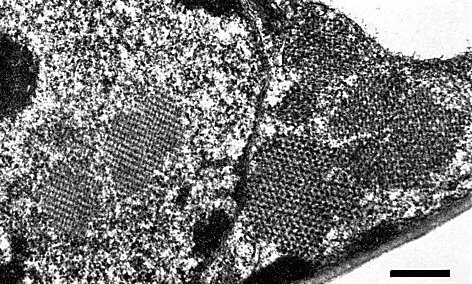
Electron micrograph of virus-like particles in nucleus and cytoplasm of Phaseolus vulgaris. Bar represents 1 µm. Photograph courtesy of D. Lesemann.
References list for DPV: Desmodium yellow mottle virus (168)
- Dunn & Hitchborn, Virology 25: 171, 1965.
- Edwardson, Purcifull, Zettler, Christie & Christie, Pl. Dis. Reptr 54: 161, 1970.
- Koenig, Virology 72: 1, 1976.
- Koenig & Givord, Virology 58: 119, 1974.
- Moore, Scott & Walters, Pl. Dis. Reptr 53: 154, 1969.
- Reichmann, Virology 25: 166, 1965.
- Scott & Moore, Virology 50: 613, 1972.
- Walters & Dodd, Phytopathology 59: 1055, 1969.
- Walters & Scott, Phytopathology 62: 125, 1972.