Details of DPV and References
DPV NO: 178 September 1977
Family: Tombusviridae
Genus: Tombusvirus
Species: Cymbidium ringspot virus | Acronym: CymRSV
Cymbidium ringspot virus
M. Hollings Glasshouse Crops Research Institute, Littlehampton, Sussex BN16 3PU, England
Olwen M. Stone Glasshouse Crops Research Institute, Littlehampton, Sussex BN16 3PU, England
Contents
- Introduction
- Main Diseases
- Geographical Distribution
- Host Range and Symptomatology
- Strains
- Transmission by Vectors
- Transmission through Seed
- Transmission by Grafting
- Transmission by Dodder
- Serology
- Nucleic Acid Hybridization
- Relationships
- Stability in Sap
- Purification
- Properties of Particles
- Particle Structure
- Particle Composition
- Properties of Infective Nucleic Acid
- Molecular Structure
- Genome Properties
- Satellite
- Relations with Cells and Tissues
- Ecology and Control
- Notes
- Acknowledgements
- Figures
- References
Introduction
-
Described by
Hollings & Stone (1963).
Synonyms
- White clover virus (Rev. appl. Mycol. 45: 74)
-
An RNA-containing virus with polyhedral particles c. 30 nm in diameter, found in cymbidium orchids and white clover (Trifolium repens) in Britain. The virus is readily sap-transmissible to a wide range of herbaceous plant species, although few are systemically infected. No vector is known, but the virus is exuded by roots of infected plants and can infect healthy plants grown in contaminated soil.
Main Diseases
In Cymbidium spp., the virus causes slight chlorotic ring-mottle (Fig. 2); it intensifies the symptoms induced by cymbidium mosaic virus when present in mixed infections. In Trifolium repens it produces chlorotic flecks and mottling with slight stunting (Fig. 1). However, the symptoms in clover and Cymbidium are not diagnostic. Although highly contagious, the virus has apparently not become widely established in clover pastures.
Geographical Distribution
Southern England.
Host Range and Symptomatology
The virus, which is readily transmitted by inoculation of sap, infected over 60 of 101 plant species tested, in 23 of 35 plant families. Most suscepts produced only local lesions, only 13 and 18 species respectively being systemically infected by the cymbidium and clover strains (Hollings et al., 1977).
-
Diagnostic species
- Chenopodium amaranticolor, C. murale
and C. quinoa. Small, chlorotic to pale fawn necrotic local lesions in 4-6 days (Fig. 3); no systemic infection. - Emilia sagittata. Chlorotic local lesions in 6-8 days; systemic yellow spots
and rings,
often becoming brown and necrotic, with leaf malformation and severe necrosis
(Fig. 5).
Symptomless invasion
of stems sometimes occurs.
- Nicotiana clevelandii. Both strains induced numerous diffuse chlorotic, and fewer brown necrotic local lesions 2-3 mm in diameter in 3-5 days, usually expanding to necrotic areas and soon killing the leaf. Systemic chlorotic and necrotic small speckles, severe puckering, stunting and rosetting follow in 7-10 days (Fig. 6). Small plants are usually killed in 2-3 weeks.
- N. glutinosa, N. tabacum (tobacco), White Burley cvs. Judy's Pride and Havana 425, and Xanthi-nc. Small, brown necrotic local lesions in 5-7 days. No systemic invasion.
- Phaseolus vulgaris (French bean) cv. The Prince. Small, whitish necrotic local lesions in 4-7 days with the cymbidium strain (Fig. 4); semi-necrotic to chlorotic local lesions with the clover strain. Both strains often become systemic, with chlorosis and slight mottle.
- Vigna sinensis (cowpea) cv. Blackeye. Necrotic whitish and brown local lesions in 3-6 days; no systemic infection.
- Nicotiana clevelandii. Both strains induced numerous diffuse chlorotic, and fewer brown necrotic local lesions 2-3 mm in diameter in 3-5 days, usually expanding to necrotic areas and soon killing the leaf. Systemic chlorotic and necrotic small speckles, severe puckering, stunting and rosetting follow in 7-10 days (Fig. 6). Small plants are usually killed in 2-3 weeks.
-
Propagation species
- Nicotiana clevelandii
gives very high virus yields; the virus is best maintained as leaves dried over anhydrous CaCl2 at 0°C, or as lyophilized sap.Assay species
- Chenopodium quinoa
and Phaseolus vulgaris are suitable for local lesion assay.
Strains
Two strains, from cymbidium and white clover respectively, have been differentiated by host range and serological tests. The cymbidium strain did not infect Trifolium incarnatum or T. repens. The clover strain caused systemic mottle in tomato (Lycopersicon esculentum) cv. Moneymaker, whereas the cymbidium strain produced only symptomless infection in the inoculated leaves (Hollings et al., 1977).
Transmission by Vectors
No transmission by the aphids Myzus persicae or Acyrthosiphon pisum. The virus was released into sterilized soil from roots of infected plants, and was readily acquired by bait seedlings planted in such soil. Infection also occurred when purified virus was applied to the surface of sterilized soil containing bait plants; there is no evidence for any living vector (Hollings et al., 1977).
Transmission through Seed
No seed transmission was detected in Trifolium incarnatum, Phaseolus vulgaris cv. The Prince, or Nicotiana clevelandii.
Serology
The virus is a good immunogen and reacts well in vitro. Antisera prepared in rabbits by one intravenous, and two intramuscular injections (with Freund’s complete adjuvant) over 3 weeks had titres against purified virus preparations in tube precipitin tests of 1/2048 to 1/4096, the precipitates being somatic (granular). In immunodiffusion, antiserum titres up to 1/512 were obtained. Microprecipitin and ring tests are also satisfactory (Hollings et al., 1977).
Relationships
The two strains were serologically fairly closely related, forming small spurs in immunodiffusion (Fig. 8). Both strains were serologically unrelated to any of 58 strains of 43 other isometric viruses, including 18 tombusvirus serotypes. The virus nevertheless shares many properties with tombusviruses (e.g. particle morphology, sedimentation coefficient, buoyant density in CsCl, M. Wt of ssRNA and coat polypeptides, precipitation in salt-free medium), and has many similarities in host range and symptoms (Hollings & Stone, 1965; Hollings et al., 1977). We therefore provisionally assign it to that group.
Stability in Sap
The virus is extremely stable. It was infective in Nicotiana clevelandii sap at dilutions to 10-5-10-6, but only to 2 x 10⁻¹ in cymbidium sap. Infectivity withstood 10 min at 85-90°C (very occasionally 95°C), although over 98% of infectivity was lost at 75-80°C; it survived 14 months at 20°C and at least 12½ years at 2°C. Lyophilized sap was highly infective after 13½ years at c. 20°C in ampoules sealed under high vacuum (Hollings et al., 1976; 1977).
Purification
The virus is easily purified from systemically infected Nicotiana clevelandii (Hollings & Stone, 1965; Hollings et al., 1977). Harvest N. clevelandii plants c. 2 weeks after infection, homogenize in a blender in 0.05 M phosphate buffer, pH 7.6 (2 ml/g tissue), containing 0.01 M EDTA and 0.14% (v/v) 2-mercaptoethanol, and filter through cloth. Add n-butanol dropwise (9.3 ml/100 ml juice) and stir for 1 h, then keep at c. 4°C for 18-24 h, and separate the virus by one or more cycles of differential centrifugation (12,000 g for 25 min; 78,000 g for 90 min). Re-suspend the final pellets in 0.03 M phosphate buffer (1 ml/30 g original leaf) and after 1-2 h remove insoluble material by brief centrifugation (10,000 g for 10 min). The virus precipitates in the absence of salts, at least 0.005 M being necessary to prevent this.
Further purification can be effected by sucrose density-gradient centrifugation and reconcentration from the virus-containing zone, but up to 90% of the virus can be lost. A much better procedure is molecular permeation chromatography on columns of controlled-pore glass beads (Barton, 1977; Hollings et al., 1977). To prevent non-specific adsorption, the column (80 x 1.5 cm) of glass beads (70 nm pore size) is pre-treated with a 1% (w/v) solution of polyethylene glycol (M. Wt 20,000) in 0.04 M phosphate buffer pH 7.0. Samples of 1-2 ml of partially purified virus are then applied to the column and eluted with 0.05 M sodium phosphate buffer pH 7.0. The virus elutes immediately after the void volume, separated from ribosomal and other host material.
Properties of Particles
Purified preparations usually produce a single light-scattering virus band in density-gradient centrifugation, but occasionally multiple bands occur (Hollings & Stone, 1965). A single sedimenting component is obtained in analytical ultracentrifugation (s°20, w = 137 S).
A260/A280: 1.55; Amax(260)/Amin(242): 1.17 (values corrected for light-scattering).
Buoyant density: 1.363 g/ml in CsCl (Hollings et al., 1977).
Immunoelectrophoresis: in 0.8% Ionagar no. 2 in 0.03 M phosphate buffer pH 7.6, both strains of the virus migrated as single antigenic components towards the cathode, at c. 54 x 10-6 cm2 sec-1 v-1 (Hollings et al., 1977).
Particle Structure
Particles are polyhedral, c. 30 nm in diameter in 2% ammonium molybdate + saturated uranyl acetate (Fig. 7), but are damaged and swollen to c. 35-40 nm by long exposure to 2% phosphotungstate (Hollings et al., 1977).
Particle Composition
Nucleic acid: RNA, single-stranded, about 15% of particle weight (estimated spectrophotometrically), and of M. Wt c. 1.7 x 10⁶ from polyacrylamide gel electrophoresis. Molar percentages of nucleotides: G 27.8 ± 1.7: A 24.9 ± 0.9: C 21.3 ± 1.7: U 26.1 ± 1.6 (Hollings et al., 1977).
Protein: one coat polypeptide of M. Wt 43,600 ± 1300.
Relations with Cells and Tissues
The virus is present in leaves, stems, flowers and roots of clover and Nicotiana clevelandii.
Notes
The virus was eliminated from 96% of small cuttings (c. 5 mm) taken from infected Nicotiana clevelandii plants grown at 35-37°C for 9 weeks.
One isolate from Trifolium repens contained two antigenic components designated A and B. A was serologically indistinguishable from other isolates from T. repens. B was unrelated to either the clover or cymbidium strains of the virus, or to any of 51 other isometric viruses. Antigen B could not be propagated alone and could not be separated from the virus by analytical centrifugation, or by isopycnic banding. Available evidence suggests that it may be a satellite virus (Hollings et al., 1976).
Figures
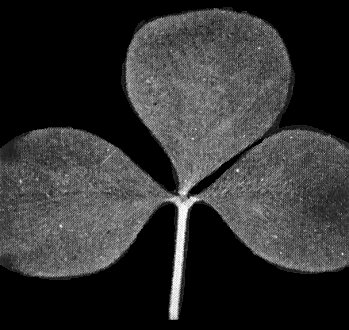
Leaf of Trifolium incarnatum systemically infected with the clover strain of the virus. Symptoms in T. repens are very similar.

Leaves of cymbidium; (left) systemically infected with the cymbidium strain of the virus; (right) healthy leaf.

Local lesions (cymbidium strain) in Chenopodium quinoa 6 days after inoculation.

Local lesions (cymbidium strain) in Phaseolus vulgaris cv. The Prince 7 days after inoculation.

Leaf of Emilia sagittata systemically infected with the cymbidium strain.
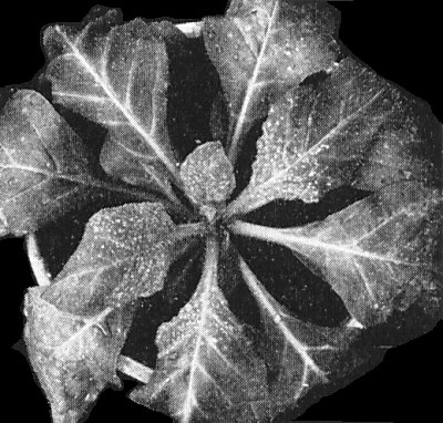
Nicotiana clevelandii plant 9 days after infection with the clover strain, showing systemic necrotic speckles, mottle and dwarfing.

Partially purified preparation of the virus (clover strain) stained in uranyl formate. Bar represents 100 nm.

Immunodiffusion plates, stained with amido black. (Upper picture) left well = clover strain; right well = cymbidium strain; upper well = antiserum to cymbidium strain. Shows spur formation between clover and cymbidium strains, and single lines of precipitation.
(Lower picture) left well = clover strain, antigen A only; right well = clover strain, antigens A + B; upper well = antiserum to clover strain with A + B components. Note the two lines of precipitation for A and B antigens respectively, with the latter (nearer to the antigen well) unrelated to antigen A. All antigens in partially purified preparations.

Soil transmission of the clover strain: N. clevelandii bait seedlings planted in contaminated soil. Infected seedling (upper) and uninfected seedling (lower), 3 weeks after planting.
References list for DPV: Cymbidium ringspot virus (178)
- Barton, J. gen. Virol. 35: 77, 1977.
- Hollings & Stone, Rep. Glasshouse Crops Res. Inst. for 1962: 90, 1963.
- Hollings & Stone, Ann. appl. Biol. 56: 87, 1965.
- Hollings, Stone & Barton, Ann. appl. Biol. 86: 233, 1977.
- Hollings, Stone & Pawley, Rep. Glasshouse Crops Res. Inst. for 1975: 120, 1976.