Details of DPV and References
DPV NO: 179 September 1977
Family: Luteoviridae
Genus: Luteovirus
Species: Soybean dwarf virus | Acronym: SbDV
Soybean dwarf virus
T. Tamada Hokkaido Central Agricultural Experiment Station, Naganuma, Hokkaido 069-13, Japan
M. Kojima Department of Botany, Faculty of Agriculture, Hokkaido University, Sapporo 060, Japan
Contents
- Introduction
- Main Diseases
- Geographical Distribution
- Host Range and Symptomatology
- Strains
- Transmission by Vectors
- Transmission through Seed
- Transmission by Grafting
- Transmission by Dodder
- Serology
- Nucleic Acid Hybridization
- Relationships
- Stability in Sap
- Purification
- Properties of Particles
- Particle Structure
- Particle Composition
- Properties of Infective Nucleic Acid
- Molecular Structure
- Genome Properties
- Satellite
- Relations with Cells and Tissues
- Ecology and Control
- Notes
- Acknowledgements
- Figures
- References
Introduction
Described by Tamada et al. (1969) and Tamada (1970).
A virus with isometric particles about 25 nm in diameter found in Japan. It is restricted to the Leguminosae and is transmitted by the aphid Acyrthosiphon (Aulacorthum) solani in the persistent (circulative) manner but not by inoculation of sap.
Main Diseases
Causes severe stunting of soybean (Glycine max). Leaves are puckered (Fig. 3) and show interveinal yellowing. The severity of symptoms depends on the soybean cultivar and virus strain. A 50% incidence of field infection may result in as much as 40% reduction in yield. Also causes a yellows disease of French bean (Phaseolus vulgaris). Only mild yellowing symptoms or none are produced in pea (Pisum sativum). Red clover (Trifolium pratense) and white clover (T. repens) are naturally infected with the virus without showing symptoms (Tamada, 1973; 1975).
Geographical Distribution
Found in Japan. Although the distribution of the virus is limited to Hokkaido and the northern areas of Honshu, the virus is spreading south at a rate of about 30 km per year.
Host Range and Symptomatology
The virus is restricted in host range to members of the Leguminosae (Tamada, 1970; 1973; 1975). Occurs naturally in a number of perennial legumes including Trifolium spp. and often in common overwintering hosts of vector aphids. In general, the symptoms of virus infection are stunting, chlorosis and interveinal yellowing or marginal reddening of older leaves. The virus is not transmitted by inoculation of sap.
- Diagnostic species
- Glycine max (soybean). The first symptoms are faint
yellowing and slight chlorosis of the youngest leaflets 1-2 weeks
after infection. With the dwarfing strain, the leaflets are reduced
in size and curled downward; the plants are stunted with shortened
petioles and internodes (Fig. 1) and the leaves later become dark
green and thickened. With the yellowing strain, older leaves show
interveinal yellowing and become brittle (Fig. 5); the leaflets may
become puckered (Fig. 3), depending on the cultivar and environmental
conditions.
- Astragalus sinicus (milk-vetch). Chlorosis and slight rolling of the leaves (Fig. 4).
- Trifolium incarnatum (crimson clover), T. dubium (suckling clover) and T. subterraneum (subterranean clover). Stunting of plants and chlorosis and marginal reddening of older leaves.
- Vicia faba (broad bean). Interveinal chlorosis of intermediate or lower leaves (Fig. 2).
- Astragalus sinicus (milk-vetch). Chlorosis and slight rolling of the leaves (Fig. 4).
- Propagation species
- Glycine max (cv. Shiro Tsurunoko) is suitable for maintaining
cultures and as a source of virus for purification.
- Assay species
- Glycine max (cv. Shiro Tsurunoko) is suitable for insect transmission tests. Two bioassay methods, injection of virus into aphids and acquisition of virus by membrane-feeding, can be useful (Tamada, 1975; Kojima & Tamada, 1976).
Strains
Two strains, designated as dwarfing and yellowing, have been identified on the basis of host range and symptoms (Tamada, 1973; 1975). Dwarfing strains infect red clover (Trifolium pratense), but not French bean (Phaseolus vulgaris), groundnut (Arachis hypogaea) or Lupinus cosenlini, whereas yellowing strains infect French bean, groundnut and L. cosenlini, but not red clover. In general, the symptoms caused by yellowing strains are more severe than those caused by dwarfing strains.
Transmission by Vectors
The virus is transmitted by the aphid Acyrthosiphon (Aulacorthum) solani in the persistent (circulative) manner, but not by six other species of aphids tested: Aphis glycines, Aphis craccivora, Myzus persicae, Acyrthosiphon pisum, Acyrthosiphon kondoi and Macrosiphum euphorbiae (Tamada et al., 1969; Tamada, 1975). Minimum acquisition access period is between 30 and 60 min and minimum inoculation access period between 10 and 30 min. The minimum latent period in the vector is beween 15 and 27 h, depending on the length of the acquisition access period (Tamada, 1970; 1975). Nymphs transmit the virus more efficiently than adults. In serial transmission tests, the aphids retain ability to transmit after moulting and for periods up to 40 days, but most of them cease to transmit in the later transfers. The virus is not transmitted to the progeny of infective aphids. Transmission efficiency of aphids can be increased by additional acquisition access (recharging). There is no evidence for multiplication of the virus within the body of the vector (Tamada, 1975).
Transmission through Seed
Not seed-transmitted.
Transmission by Dodder
Not tested.
Serology
The virus is strongly immunogenic, but concentrated or partially purified virus preparations must be used in all serological tests, because of the low concentration in the plants. Antisera with titres of 1/2048-1/4096 (ring precipitin test) were obtained by intramuscular and intravenous injection of rabbits (Tamada, 1975; Kojima & Tamada, 1976). The virus reacts in micro agar-gel double diffusion tests, diffusing to produce a single precipitin line. Infectivity neutralization tests, demonstrated by feeding aphids through membranes on virus-antiserum mixtures, have also been successful.
Relationships
No serological differences were detected between dwarfing and yellowing strains (Tamada, 1975; Kojima & Tamada, 1976), although no protection was found between them in soybean plants (Tamada, 1973). Mild isolates (Fig. 1) of the dwarfing strain gave interference against more virulent isolates inoculated by aphids (Tamada, 1973).
In mode of transmission, symptomatology, particle morphology and virus localization within the phloem, soybean dwarf virus resembles members of the luteovirus group which includes barley yellow dwarf virus (Rochow, 1970) and beet western yellows virus (Duffus, 1972). Using infectivity neutralization tests, Duffus (1977) detected serological relationships among soybean dwarf virus, beet western yellows virus, turnip yellows virus, beet mild yellowing virus, and the RPV strain of barley yellow dwarf virus but in gel-diffusion tests (T. Tamada, unpublished data) soybean dwarf virus did not react with antisera prepared against beet western yellows and turnip yellows viruses. No serological relationship was detected between potato leafroll virus and either soybean dwarf virus (Murayama & Kojima, 1974; Kojima & Tamada, 1976) or beet western yellows virus (Duffus & Gold, 1969).
Stability in Sap
Determined for both strains of the virus using concentrated or partially purified virus preparations, and assaying by insect injection and membrane-feeding methods. Thermal inactivation point (10 min) is between 45 and 50°C; dilution end-point of unconcentrated sap, 1/2; infectivity survives for at least 4 months at 4°C, and for at least 20 days at 15°C. Infectivity of virus is not affected by three cycles of freezing and thawing (Tamada, 1975; Kojima & Tamada, 1976).
Purification
The virus can be purified from infected soybean plants by the following method (Kojima & Tamada, 1976). Grind fresh plants in 0.5 M phosphate buffer, pH 7.4, containing 0.01 M disodium ethylenediamine-tetraacetate (EDTA). Clarify the extracts with one-half volume of a 1:1 mixture of chloroform and n-butanol and precipitate the virus by adding polyethylene glycol (PEG, M. Wt 6000) to 8%. Re-clarify the resuspended preparations with one-half volume of Diafron S-3 (trifluorotrichloroethane) and purify the virus further by differential centrifugation and sucrose density gradient centrifugation. Resuspend all pellets in 0.01 M phosphate buffer, pH 7.4, containing 0.001 M EDTA. Virus yields are 100-400 µg per kg of tissue.
The following modification of the procedure is also used. Homogenize frozen tissue in 2-3 volumes of 0.1 M phosphate buffer, pH 7.4, containing 0.5% 2-mercaptoethanol. Express juice through cheesecloth and clarify the extracts with a 1:1 mixture of chloroform and n-butanol, and precipitate the virus by adding PEG to 8%. Resuspend the pellets in 0.01 M phosphate buffer containing 1.0% Triton X-100. Purify the virus further by centrifugation through a 20% sucrose cushion and then by repeated sucrose density gradient centrifugation. A good increase in virus yield is obtained by grinding the fibres retained by cheesecloth in liquid nitrogen.
Properties of Particles
Virus preparations contain a single sedimenting component.
A260/A280: 1.96 (dwarfing strain), 1.90 (yellowing strain) (not corrected for light-scattering) (Kojima & Tamada, 1976).
Particle Structure
Particles of both strains are isometric, about 25 nm in diameter in negatively stained preparations (Fig. 6). The particles present hexagonal profiles with some surface structure.
Particle Composition
Nucleic acid: No information.
Protein: Subunit M. Wt (estimated by
SDS-polyacrylamide gel electrophoresis) about 22,000 (Kojima &
Shikata, 1976).
Relations with Cells and Tissues
Virus particles are found in the sieve tubes, phloem parenchyma and companion cells, and xylem vessels. The virus particles are associated with phloem necrosis; the dwarfing strain seems to cause more severe degeneration of phloem tissues than the yellowing strain (Tamada, 1975).
Notes
Soybean dwarf virus resembles some other persistent (circulative) aphid-borne viruses of legumes including bean leafroll, subterranean clover stunt, milk-vetch dwarf, and subterranean clover red leaf viruses. These viruses may be related but serological evidence is not available. However, soybean dwarf virus can be distinguished from the other four viruses by vector species, host range and symptomatology, as follows:
1. Bean leafroll virus (synonymous with pea leafroll, pea tip yellowing,
pea top yellows, and pea yellows viruses) which causes yellowing and
leaf-rolling in pea, field bean and vetch in Europe (Quantz &
Völk, 1954; Thottappilly, 1969; Cockbain
& Gibbs, 1973), is transmitted by Acyrthosiphon pisum,
Macrosiphum euphorbiae and Myzus persicae, and infects
Lathyrus odoratus, Medicago sativa and Vicia villosa
but not Trifolium alexandrinum and T. hybridum.
2. Subterranean clover stunt virus, which causes stunting in
subterranean clover and French bean in Australia (Grylls &
Butler, 1959; Smith, 1966), is transmitted by Aphis craccivora,
Macrosiphum euphorbiae and Myzus persicae, and infects Dolichos lablab, Medicago sativa, Melilotus alba and
Vigna sinensis but not T. hybridum.
3. Milk-vetch dwarf virus, which causes stunting and yellowing
in milk-vetch, pea and broad bean in Japan (Matsuura, 1953; Inouye
et al., 1968), is transmitted by Aphis craccivora
but not by Acyrthosiphon (Aulacorthum) solani, and infects Lathyrus odoratus, Medicago sativa, Melilotus alba,
Vigna sinensis and several non-legumes but not Trifolium
hybridum, T. pratense and T. repens.
4. Subterranean clover red leaf virus, which causes red leaf in
subterranean clover, and yellowing and leaf rolling in pea, French
bean, broad bean and lupin in New Zealand (Wilson & Close, 1973;
R. C .Close, personal communication), resembles soybean
dwarf virus in its transmission by Acyrthosiphon (Aulacorthum)
solani, and in its symptoms on soybean plants, but infects
several non-leguminous hosts including Rumex obtusifolius,
Erodium moschatum and E. cicutarium
(J. W. Ashby, R. C. Close & P. B. Teh, unpublished data).
Beet western yellows virus also induces yellowing, leaf rolling and stunting of various legumes which include Cicer arietinum, Lathyrus odoratus, Pisum sativum, Trifolium alexandrinum, T. incarnatum, Vicia faba and Glycine max (Duffus, 1960; 1964; Duffus & Milbrath, 1977; Duffus & Russell, 1970), but it differs from soybean dwarf virus in having a very wide range of hosts.
The name soybean dwarf virus may be confused with soybean stunt virus, but these viruses are quite different, the latter being a strain of cucumber mosaic virus (Takahashi et al., 1970).
Although none of the soybean varieties tested are immune to the virus, the use of tolerant varieties is an important way of controlling soybean dwarf disease. The use of systemic insecticides can also reduce virus spread within fields (Tamada, 1975).
Figures
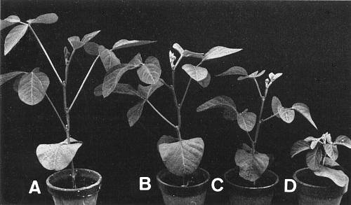
Soybean plants (cv. Shiro Tsurunoko) infected with isolates of the dwarfing strain, showing various degrees of dwarfing. A-healthy, B-mild isolate (SDV-DM), C-intermediate symptom caused by mixed infection of mild isolate (SDV-DM) and severe isolate (SDV-DS), D-severe isolate (SDV-DS).

Interveinal chlorosis in intermediate leaves of broad bean infected with the yellowing strain (SDV-Y).

Stunting and rugosity in naturally infected soybean plants (cv. Oshimashirome).
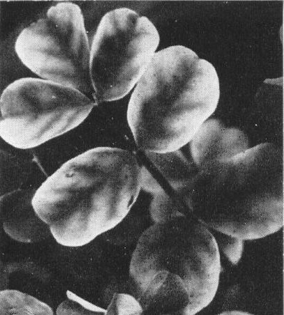
Chlorosis and rolling in leaf of milk-vetch infected with the yellowing strain (SDV-Y).

Interveinal yellowing in the older leaflet of soybean (cv. Gokuwasechishima) infected with the yellowing strain (SDV-Y).

Virus particles from a purified preparation stained in 2% phosphotungstate. Bar represents 100 nm.
References list for DPV: Soybean dwarf virus (179)
- Cockbain & Gibbs, Ann. appl. Biol. 73: 177, 1973.
- Duffus, Phytopathology 50: 389, 1960.
- Duffus, Phytopathology 54: 736, 1964.
- Duffus, CMI/AAB Descriptions of Plant Viruses 89, 4pp., 1972.
- Duffus, Phytopathology 67: 1197, 1977.
- Duffus & Gold, Virology 37: 150, 1969.
- Duffus & Milbrath, Phytopathology 67: 269, 1977.
- Duffus & Russell, Phytopathology 60: 1199, 1970.
- Grylls & Butler, Aust. J. agric. Res. 10: 145, 1959.
- Inouye, Inouye & Mitsuhata, Ann. phytopath. Soc. Japan 34: 28, 1968.
- Kojima & Shikata, Ann. phytopath. Soc. Japan 42: 92, (abstr.), 1976.
- Kojima & Tamada, Phytopath. Z. 85: 237, 1976.
- Matsuura, Ann. phytopath. Soc. Japan 17: 65, 1953.
- Murayama & Kojima, Proc. Japan Acad. 50: 322, 1974.
- Quantz & Völk, NachrBl. dt. PflSchutzdienst., Stuttg. 6: 177, 1954.
- Rochow, CMI/AAB Descriptions of Plant Viruses 32, 4 pp., 1970.
- Smith, Aust. J. agric. Res. 17: 875, 1966.
- Takahashi, Utagawa, Tomaru & Saito, Ann. phytopath. Soc. Japan 36: 374 (abstr.), 1970.
- Tamada, Ann. phytopath. Soc. Japan 36: 266, 1970.
- Tamada, Ann. phytopath. Soc. Japan 39: 27, 1973.
- Tamada, Rep. Hokkaido prefec. agric. Exp. Stn 25, 144 pp., 1975.
- Tamada, Goto, Chiba & Suwa, Ann. phytopath. Soc. Japan 35: 282, 1969.
- Thottappilly, Z. PflKrankh. PflPath. PflSchutz. 76: 67, 1969.
- Wilson & Close, N.Z. Jl agric. Res. 16: 305, 1973.