Details of DPV and References
DPV NO: 224 September 1980
Family: Unallocated ssRNA+ viruses
Genus: Sobemovirus
Species: Lucerne transient streak virus | Acronym: LTSV
Lucerne transient streak virus
R. L. S. Forster Scottish Horticultural Research Institute, Invergowrie, Dundee, Scotland
A. T. Jones Scottish Horticultural Research Institute, Invergowrie, Dundee, Scotland
Contents
- Introduction
- Main Diseases
- Geographical Distribution
- Host Range and Symptomatology
- Strains
- Transmission by Vectors
- Transmission through Seed
- Transmission by Grafting
- Transmission by Dodder
- Serology
- Nucleic Acid Hybridization
- Relationships
- Stability in Sap
- Purification
- Properties of Particles
- Particle Structure
- Particle Composition
- Properties of Infective Nucleic Acid
- Molecular Structure
- Genome Properties
- Satellite
- Relations with Cells and Tissues
- Ecology and Control
- Notes
- Acknowledgements
- Figures
- References
Introduction
Described by Blackstock (1974, 1978) and Forster & Jones (1979).
A virus with RNA-containing isometric particles 27-28 nm in diameter, which is manually transmissible to species in at least four plant families. Its means of natural spread is not known. Reported to occur commonly in lucerne crops in Australia and New Zealand.
Main Diseases
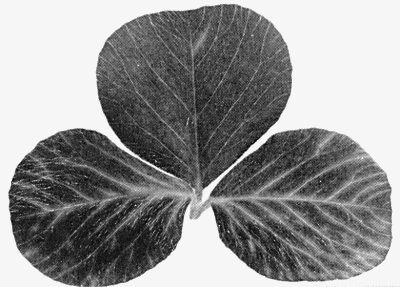
In lucerne, the virus causes chlorotic streaking along the lateral veins and sometimes distortion of the leaves (Fig. 1). Symptoms may persist but are often transient or absent. In a field trial of infected lucerne, Blackstock (1978) reported a loss of 18% in dry matter yield.
Geographical Distribution
Reported only from lucerne (Medicago sativa) in Australia and New Zealand.
Host Range and Symptomatology
The virus has a moderately wide host range, infecting 18 species from four families. It is readily transmitted to most hosts by inoculation with sap but transmission to lucerne is difficult unless a purified virus preparation, or sap from infected Trifolium incarnatum, is used as inoculum (Blackstock, 1978; Forster & Jones, 1979). Some differences in host range and symptomatology are reported between Australian and New Zealand isolates but the isolates from the two sources have not been compared directly.
-
Diagnostic species
- Chenopodium amaranticolor.
Pin-point necrotic local lesions in 3-5 days (Fig. 4). Isolates from Australia induce systemic chlorotic blotches whereas isolates from New Zealand do not infect systemically. - Chenopodium quinoa. Chlorotic or necrotic local lesions in 3-4 days.
Some plants develop
scattered systemic chlorotic blotches.
- Nicotiana clevelandii. New Zealand isolates induce large chlorotic local lesions and occasional systemic chlorotic mottle (Fig. 2). Not infected by Australian isolates.
- Pisum sativum (pea) cvs Greenfeast, Onward. New Zealand isolates induce local necrotic rings (Fig. 5) but Australian isolates infect symptomlessly; not systemic.
- Trifolium incarnatum (crimson clover). Persistent yellow flecks and streaks develop along the lateral veins of systemically infected leaves (Fig. 3).
- Nicotiana clevelandii. New Zealand isolates induce large chlorotic local lesions and occasional systemic chlorotic mottle (Fig. 2). Not infected by Australian isolates.
-
Propagation species
- The virus may be propagated for purification in Chenopodium quinoa and Nicotiana clevelandii. Lucerne and crimson clover are suitable for maintaining cultures.
-
Assay species
- Chenopodium amaranticolor
and C. quinoa.
Strains
All New Zealand isolates tested are indistinguishable serologically, and in host range and symptomatology (Forster & Jones, 1979). Australian and New Zealand isolates appear to differ in host range and symptomatology (Blackstock, 1978; Forster & Jones, 1979) but isolates from the two countries are closely related serologically.
Transmission by Vectors
No vectors reported. The aphids Acyrthosiphon kondoi, A. solani, Aphis craccivora, Myzus ornatus and M. persicae failed to transmit the virus (Blackstock, 1978; Forster & Jones, 1979) and no transmission occurred in limited soil transmission tests (Blackstock, 1978).
Transmission through Seed
No seed transmission was detected in Chenopodium quinoa or lucerne in Australia (Blackstock, 1978). In New Zealand, no seed transmission was detected in seedlings from four commercial lucerne seed lines, two of which contained Australian lucerne latent virus (Forster & Jones, 1979; Jones, Forster & Mohamed, 1979).
Transmission by Dodder
The virus was not transmitted by Cuscuta campestris from lucerne to lucerne (R. L. S. Forster, unpublished data).
Serology
The virus is weakly immunogenic, giving antisera with titres of 1/128-1/512 in gel-diffusion tests. Stabilization of the virus immunogen with formaldehyde did not increase the antiserum titre (Blackstock, 1978). New Zealand isolates are readily detected in infective sap of Nicotiana clevelandii and Chenopodium quinoa by gel-diffusion tests.
Relationships
Particle morphology, sedimentation behaviour in CsCl and Cs2SO4, lability in EDTA at alkaline pH, and the M. Wt of the coat protein and the single RNA species, suggest affinities to the proposed southern bean mosaic virus group (Hull, 1977). The virus is distinguishable from other reported members of this group in host range, symptomatology and serological properties (Forster & Jones, 1979). The virus failed to react with antisera to 40 distinct isometric plant viruses including the five viruses assigned to the southern bean mosaic virus group (Hull, 1977), namely cocksfoot mottle, rice yellow mottle, southern bean mosaic, sowbane mosaic and turnip rosette viruses.
Stability in Sap
In N. clevelandii sap, the infectivity of a New Zealand isolate was retained after dilution 10-5 but not 10-6, after heating for 10 min at 70° but not 75°C, and after storage for 4 but not 5 weeks at room temperature (Forster & Jones, 1979).
Purification
The virus is relatively stable and can be readily purified from C. quinoa and N. clevelandii by clarification with 8.5% n-butanol or 50% chloroform, differential centrifugation and sucrose density gradient centrifugation (Blackstock, 1978; Forster & Jones, 1979).
Properties of Particles
Purified preparations contain a single infective sedimenting component. A New Zealand isolate contained a single electrophoretic component which migrated towards the cathode in 0.025 M sodium phosphate buffered agarose at pH 7 and 8, and formed one buoyant density component in CsCl but two components in Cs2SO4.
A260/A280: 1.52.
Absorbance at 260 nm (1 mg/ml, 1 cm light path): 5.2.
Buoyant density in CsCl: 1.37 g/cm3.
Buoyant density in Cs2SO4: 1.25-1.28 and 1.31-1.34 g/cm3.
Sedimentation coefficient (s°20,w): 112-114 S.
Stability: Particles are degraded in 1 M CaCl2 (Blackstock, 1978). Particles are completely degraded in 10 mM EDTA + 1 M NaCl at pH 7 and 8 but not at pH 5. In 10 mM EDTA without NaCl, the particles are partially degraded at pH 8 into two sedimenting components of 92 S and 105 S (Forster & Jones, 1979).
Particle Structure
In 2% potassium phosphotungstate, pH 7, many particles are disrupted but they remain intact at pH 4.0 or in uranyl salts. In most preparations some particles are partially or completely penetrated by the stain. Unpenetrated particles are isometric, 27-28 nm in diameter, with a hexagonal outline (Fig. 6). Ring-like morphological subunits were observed on some particles after freeze-dehydration (Forster & Jones, 1979).
Particle Composition
Nucleic acid: A single infective RNA species, probably single-stranded, with a sedimentation coefficient of 26 S and M. Wt of c. 1.4 x 10⁶, estimated by polyacrylamide gel electrophoresis under non-denaturing conditions. About 18% of particle weight (estimated from phosphorus content of 1.76% by weight). Minor RNA species are sometimes present (Forster & Jones, 1979). No evidence was obtained for polyadenylate sequences but the infectivity of RNA preparations is abolished by incubation with proteinase K (M. A. Mayo & A. T. Jones, unpublished data).
Protein: One major species of M. Wt c. 32,000 estimated by polyacrylamide gel electrophoresis. A minor component of M. Wt 29,000 is present in many preparations (Forster & Jones, 1979).
Relations with Cells and Tissues
Virus-like particles occur in the cytoplasm and nucleus of cells of infected crimson clover plants (Forster & Jones, 1979).
Notes
The virus is distinguishable from other mechanically transmitted isometric viruses reported in lucerne by host range, symptomatology, sedimentation properties, serology and transmission. Thus alfalfa mosaic, cucumber mosaic and pea enation mosaic viruses are aphid transmitted, and Australian lucerne latent, tobacco ringspot, tobacco streak and tomato black ring viruses have multiple sedimenting components. The virus is not serologically related to any of these viruses.
Acknowledgements
Photographs: Plant Disease Division, Department of Scientific and Industrial Research, Auckland, New Zealand, except Fig. 6 which is courtesy of Scottish Horticultural Research Institute, Dundee.
Figures

Systemic chlorotic streaks and leaf distortion in lucerne (Medicago sativa).

Systemic chlorotic mottle in Nicotiana clevelandii.
Systemic veinal streaks and flecks in crimson clover (Trifolium incarnatum).

Pin-point necrotic local lesions in Chenopodium amaranticolor.

Local necrotic rings in pea (Pisum sativum) cv. Onward.

Virus particles negatively stained in 2% uranyl acetate, pH 3.5, after freeze-dehydration. Bar represents 100 nm.
References list for DPV: Lucerne transient streak virus (224)
- Blackstock, Newsl. Aust. Pl. Path. Soc. No. 3: 6, 1974.
- Blackstock, Aust. J. agric. Res. 29: 291, 1978.
- Forster & Jones, Ann. appl. Biol. 93: 181, 1979.
- Hull, J. gen. Virol. 36: 289, 1977.
- Jones, Forster & Mohamed, Ann. appl. Biol. 92: 49, 1979.