Details of DPV and References
DPV NO: 279 July 1984
Family: Tymoviridae
Genus: Tymovirus
Species: Voandzeia necrotic mosaic virus | Acronym: VNMV
Voandzeia necrotic mosaic virus
C. Fauquet Laboratoire de Virologie, Centre ORSTOM d'Adiopodoumé, B.P. V.51, Abidjan, Côte d'lvoire
A. Monsarrat Laboratoire de Virologie, Centre ORSTOM d'Adiopodoumé, B.P. V.51, Abidjan, Côte d'lvoire
J. C. Thouvenel Laboratoire de Virologie, Centre ORSTOM d'Adiopodoumé, B.P. V.51, Abidjan, Côte d'lvoire
Contents
- Introduction
- Main Diseases
- Geographical Distribution
- Host Range and Symptomatology
- Strains
- Transmission by Vectors
- Transmission through Seed
- Transmission by Grafting
- Transmission by Dodder
- Serology
- Nucleic Acid Hybridization
- Relationships
- Stability in Sap
- Purification
- Properties of Particles
- Particle Structure
- Particle Composition
- Properties of Infective Nucleic Acid
- Molecular Structure
- Genome Properties
- Satellite
- Relations with Cells and Tissues
- Ecology and Control
- Notes
- Acknowledgements
- Figures
- References
Introduction
-
Described by
Fauquet, Monsarrat & Thouvenel (1981) and
Monsarrat, Fauquet & Thouvenel (1984).
A virus with RNA-containing isometric particles c. 28 nm in diameter which sediment as two components. It is found in nature only in Voandzeia subterranea. Its experimental host range is restricted almost entirely to the Leguminosae. It is transmissible by inoculation with sap but not through seed. No vector is known. The virus occurs in the Ivory Coast and Upper Volta.
Main Diseases
Bambarra groundnut (Voandzeia subterranea) is the only natural host found. The virus causes stunting, mosaic, necrosis, distortion (Fig. 1, Fig. 2, Fig. 3) and sometimes the death of the plant (Monsarrat et al., 1984).
Geographical Distribution
Found occasionally in fields of Bambarra groundnut in northern Ivory Coast and southern Upper Volta.
Host Range and Symptomatology
Transmitted from Voandzeia to Voandzeia by inoculation with sap: 10-100% of plants become infected. Best results were obtained by using inocula prepared in 0.1 M borate buffer, pH 8.8, containing 0.02 M cysteine hydrochloride, 0.05 M sodium bisulphite and 0.5% bentonite (Monsarrat et al., 1984). Among 87 species tested from the families Amaranthaceae, Chenopodiaceae, Cruciferae, Cucurbitaceae, Leguminosae, Malvaceae, Passifloraceae, Scrophulariaceae and Solanaceae, only Chenopodium amaranticolor, Dolichos lablab, Vigna sinensis and V. unguiculata were infected (Monsarrat et al., 1984).
-
Diagnostic species
- Chenopodium amaranticolor.
Local necrotic lesions, developing later into red ringspots with yellow discoloration of the entire leaf. - Dolichos lablab. Chlorotic local lesions and systemic mosaic.
- Vigna unguiculata subspp. unguiculata and sinensis (cowpea). Chlorotic local lesions and systemic mosaic.
- Voandzeia subterranea (Bambarra groundnut). No local symptoms, but the plants are stunted and fail to develop. Systemically infected leaves show a yellow mosaic (Fig. 2) with necrosis along the veins leading to distortion of the leaflets (Fig. 3). Plants infected young may be killed.
- Vigna unguiculata subspp. unguiculata and sinensis (cowpea). Chlorotic local lesions and systemic mosaic.
-
Propagation species
- Voandzeia subterranea
is a good source of virus.Assay species
- Chenopodium amaranticolor
is a useful local lesion host.
Strains
None found.
Transmission by Vectors
No vector reported. The okra leaf beetle, Podagrica decolorata, and the groundnut aphid, Aphis craccivora, did not transmit the virus (Monsarrat et al., 1984).
Transmission through Seed
Not seed-borne in Voandzeia subterranea.
Serology
Very immunogenic in rabbits. Antisera with titres of 1/512-1/4096 in gel diffusion tests have been prepared.
Relationships
Voandzeia necrotic mosaic virus has many properties typical of tymoviruses including particle morphology, sedimentation behaviour, coat protein M. Wt, high thermal inactivation point, high concentration of particles in host plants, typical effects on chloroplasts and serological relationship to other members of the group. In agar gel double-diffusion tests, particles of the virus react with antisera to only four members of the group. The virus is related closely to kennedya yellow mosaic virus (SDI = 1) and distantly to turnip yellow mosaic virus (SDI = 6), desmodium yellow mottle virus (SDI = 6) and okra mosaic virus (SDI = 5) (Fauquet et al., 1981). The interrelationships among these five viruses can be expressed diagrammatically as a second ‘loop’ structure similar to that proposed by Koenig (1976) for other members of the group.
Stability in Sap
When assayed in Chenopodium amaranticolor, the virus, in crude sap of Voandzeia subterranea, loses infectivity after dilution 10-2, after heating for 10 min at 70°C, and after storage for less than 2 days at 20°C or for more than 10 days at 4°C or -20°C. Infectivity survives for several months in frozen or dried leaves.
Purification
(Monsarrat et al., 1984). Homogenise infected Voandzeia leaves in 0.2 M phosphate buffer at pH 7, containing 0.4% thioglycollic acid (3 ml/g tissue). Clarify by adding an equal volume of chloroform and concentrate by ultracentrifugation, resuspending the pellets in 0.01 M phosphate buffer, pH 7. Centrifuge the virus particles through a 20% sucrose ‘cushion’, resuspending the pellet in the same buffer. The components may be separated in sucrose density gradients. Yield is about 200 mg virus per kg infected leaves.
Properties of Particles
The particles sediment as two components in sucrose gradients, a fast sedimenting (B) component consisting of infective nucleoprotein particles, and a slower sedimenting (T) component consisting of protein shells. The two components are serologically indistinguishable.
Sedimentation coefficient (s20, w) at infinite dilution: 51 S (T), 113 S (B).
Isoelectric point: pH 4.65 ± 0.10 (B).
A260/A280: 0.64 (T), 1.75 (B).
Amax: 280 nm (T), 260 nm (B). Amin: 250 nm (T), 242 nm (B).
Amax/Amin: 1.91 (T), 1.35 (B).
Buoyant densities in CsCl (g/cm3): 1.29 (T), 1.45 (B).
Particle Structure
Particles are about 28 nm in diameter (Fig. 4) and have icosahedral symmetry. T component particles are penetrated by uranyl acetate (2%), whereas B component particles are not.
Particle Composition
Nucleic acid: Probably RNA, single-stranded, comprising more than 30% of the weight of the B particles (estimated from the absorption spectrum).
Protein: Polyacrylamide gel electrophoresis of coat protein reveals one polypeptide of M. Wt c. 20,000 ± 500.
Relations with Cells and Tissues
Crystals or high concentrations of virus-like particles occur in the cytoplasm (Fig. 5) and in the vacuoles of cells of infected Voandzeia subterranea. Peripheral vesicles bounded by a double membrane occur in the chloroplasts, as shown for other tymoviruses (Fig. 6).
Notes
Voandzeia necrotic mosaic virus is the first tymovirus to be found naturally infecting Voandzeia subterranea. Among the four tymoviruses serologically related to it, okra mosaic virus, desmodium yellow mottle virus and kennedya yellow mosaic virus have appreciably wider host ranges, whereas turnip yellow mosaic virus infects only species in the Cruciferae.
Although voandzeia necrotic mosaic virus is closely serologically related to kennedya yellow mosaic virus, the host ranges of the two viruses are completely different. Also, the characteristic red local lesions induced by voandzeia necrotic mosaic virus in Chenopodium amaranticolor are unlike those induced by other tymoviruses.
Figures
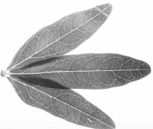
Healthy leaves of Voandzeia subterranea.
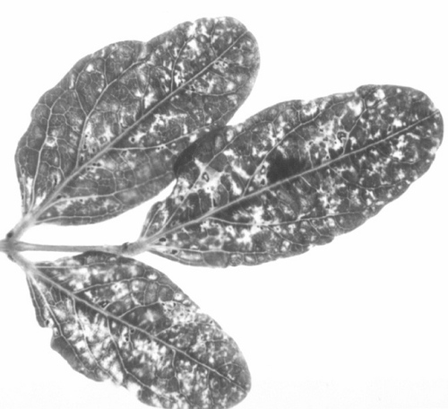
Systemically infected leaves of Voandzeia subterranea, showing mosaic.
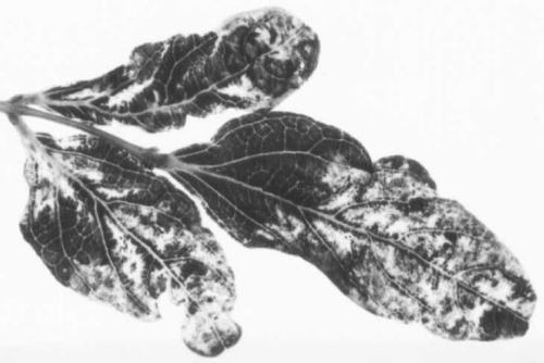
Systemically infected leaves of Voandzeia subterranea, showing necrosis.

B component particles in uranyl acetate. Bar represents 100 nm.

Virus particles aggregated on membranes in the cytoplasm of a V. subterranea leaf cell. Bar represents 100 nm.

Ultrathin section of infected V. subterranea leaf showing vesicles at different stages bounded by double membranes at the periphery of a chloroplast. Bar represents 500 nm.
References list for DPV: Voandzeia necrotic mosaic virus (279)
- Fauquet, Monsarrat & Thouvenel, Abstr. 5th Congr. Virology, Strasbourg, France, 1981: 237, 1981.
- Koenig, Virology 72: 1, 1976.
- Monsarrat, Fauquet & Thouvenel, C. r. hebd. Seanc. Acad. Sci., Paris Ser. 3, 299(3): 53, 1984.