Details of DPV and References
DPV NO: 285 July 1984
Family: Secoviridae
Genus: Cheravirus
Species: Artichoke vein banding virus | Acronym: AVBV
Artichoke vein banding virus
D. Gallitelli Dipartimento di Patologia vegetale, Università di Bari, 70126 Bari, Italy
G. P. Martelli Dipartimento di Patologia vegetale, Università di Bari, 70126 Bari, Italy
G. L. Rana Dipartimento di Patologia vegetale, Università di Bari, 70126 Bari, Italy
Contents
- Introduction
- Main Diseases
- Geographical Distribution
- Host Range and Symptomatology
- Strains
- Transmission by Vectors
- Transmission through Seed
- Transmission by Grafting
- Transmission by Dodder
- Serology
- Nucleic Acid Hybridization
- Relationships
- Stability in Sap
- Purification
- Properties of Particles
- Particle Structure
- Particle Composition
- Properties of Infective Nucleic Acid
- Molecular Structure
- Genome Properties
- Satellite
- Relations with Cells and Tissues
- Ecology and Control
- Notes
- Acknowledgements
- Figures
- References
Introduction
Described by Gallitelli, Rana & Di Franco (1978).
A virus with isometric particles c. 30 nm in diameter which sediment as three components and contain two functional species of single-stranded RNA. Readily transmitted by inoculation of sap to a moderately wide range of herbaceous hosts. The natural means of spread is unknown. Reported only from Southern Italy.
Main Diseases
Natural infection has been recorded only in artichoke (Cynara scolymus), some cultivars of which (e.g. cv. Bayrampasa) may show chlorotic discolorations along the veins (Fig. 1).
Geographical Distribution
Recorded only in Apulia (Southern Italy) in three artichoke cultivars originating from Turkey (cvs Bayrampasa and Sakiz) and Central Italy (cv. Mazzaferrata).
Host Range and Symptomatology
Experimentally the virus infected 20 species out of 71 in seven dicotyledonous families (Gallitelli et al., 1978).
- Diagnostic species
- Chenopodium quinoa and C. amaranticolor. Small
chlorotic/necrotic local lesions (Fig. 2) followed by severe systemic
mosaic and apical necrosis.
- Phaseolus vulgaris (French bean), several cvs. Pin-point necrotic local lesions; systemic mosaic, distortion and puckering of trifoliolate leaves (Fig. 3).
- Phaseolus vulgaris (French bean), several cvs. Pin-point necrotic local lesions; systemic mosaic, distortion and puckering of trifoliolate leaves (Fig. 3).
- Propagation species
- C. quinoa and P. vulgaris are good sources of virus for
purification and are suitable for maintaining cultures.
- Assay species
- C. quinoa is a satisfactory local lesion host.
Strains
None detected.
Transmission by Vectors
No information.
Transmission through Seed
No information.
Serology
The virus is a good immunogen. An antiserum with a titre of 1/256 was obtained by intramuscular and intravenous injections of rabbits with B nucleoprotein fractionated in sucrose density gradients. In gel diffusion tests this antiserum reacts equally well with B nucleoprotein and unfractionated virus, forming a single precipitin band in both instances (Gallitelli et al., 1978). Immunosorbent electron microscopy and antibody coating can be used for virus identification.
Relationships
On the basis of particle size and morphology, physico-chemical properties and intracellular behaviour the virus is classified as a tentative member of the nepovirus group (Gallitelli et al., 1978; Murant, 1981). However, it has no known vector and it was serologically unrelated to any of 24 different viruses with isometric particles including the following 17 definitive or tentative nepoviruses: arabis mosaic, artichoke Italian latent, artichoke yellow ringspot, cherry leaf roll, cherry rasp leaf, chicory yellow mottle, cocoa necrosis, grapevine Bulgarian latent, grapevine chrome mosaic, grapevine fanleaf, myrobalan latent ringspot, olive latent ringspot, peach rosette mosaic, raspberry ringspot, strawberry latent ringspot, tobacco ringspot and tomato ringspot (Gallitelli et al., 1978).
The coat protein composition of the virus too is anomalous: three small
protein species of 22,000-27,000 M. Wt instead of one large protein species
of M. Wt c. 60,000. In this respect it resembles the tentative
nepovirus, cherry rasp leaf (Stace-Smith & Hansen, 1976). Also it
resembles two definitive nepoviruses, tobacco ringspot (Chu & Francki,
1979) and olive latent ringspot (Savino, Gallitelli & Barba, 1983),
in yielding a polypeptide of M. Wt c. 15,000 after exposure to
strong denaturing conditions. If the virus is placed in the nepovirus group,
the M. Wt of its RNA-2 molecules and the sedimentation behaviour of its
nucleoprotein particles would place it in the tobacco ringspot virus sub-group
of Martelli et al. (1978) which corresponds to cluster 1 of Murant &
Taylor (1978).
Stability in Sap
In expressed sap of French bean, infectivity is lost after dilution to 10-5, heating for 10 min at 55°C or storing for 3 days at 22-24°C (Gallitelli et al., 1978).
Purification
(Gallitelli et al., 1978). Harvest infected French bean plants 10-12 days after inoculation. Homogenise each 100 g tissue in 200 ml neutral phosphate buffer containing 0.1% thioglycollic acid, squeeze homogenate through cheesecloth and clarify filtrate by the slow addition, while stirring, of 10-12% (v:v) suspension of Mg-activated bentonite (Dunn & Hitchborn, 1965). Concentrate virus by 2-3 cycles of alternate low speed (10,000 g for 10 min and high speed (78,000 g for 2 h) centrifugation and further purify by centrifuging in 10-40% sucrose gradients for 3 h at 22,000 rev/min in a Beckman SW 25.1 rotor. Average yield of virus is 1-1.5 mg/100 g infected tissue.
Properties of Particles
In sucrose density gradient and analytical ultracentrifugation (Fig. 4) purified virus preparations separate into three components (T, M and B) sedimenting at different rates. Virus particles are unstable in CsCl even after fixation for 45 min in 8% formaldehyde (Gallitelli et al., 1978).
Sedimentation coefficients, s20,w: 56 S (T), 92 S
(M), 124 S (B).
A260/A280: 1.67 (unfractionated
preparation not corrected for light-scattering).
Particle Structure
Particles are isometric, c. 30 nm in diameter, and show angular outlines. Details of surface structure are not resolved. T particles are penetrated by negative stain (Fig. 5).
Particle Composition
Nucleic acid: Single-stranded RNA comprising 24% (M) and 37% (B) of the particle weight when calculated according to Reichmann’s (1965) formula. In polyacrylamide gel electrophoresis, RNA migrates as two species (RNA-1 and RNA-2) with estimated M. Wt (x 10-6) of 2.4 and 1.4 respectively, under non-denaturing conditions (Bishop, Claybrook & Spiegelman, 1967). Both RNA species are necessary for infectivity. Whereas RNA- 1 is extracted from component B only, RNA-2 can be extracted from both M and B components (Gallitelli et al., 1978).
Protein: In SDS-polyacrylamide slab gels in the discontinuous buffer system of Laemmli (1970), protein preparations from unfractionated virus contained three polypeptides with estimated M. Wt of 22,000, 24,000 and 27,000 (Gallitelli et al., 1978). However, following dissociation under stronger denaturing conditions, i.e. 1% SDS, 2% mercaptoethanol and 6 M urea (Chu & Francki, 1979), an additional polypeptide of M. Wt 14,300 was found (Savino et al., 1983).
Relations with Cells and Tissues
The virus is present in foliar parenchyma tissues. Cytopathological modifications consist of cytoplasmic inclusion bodies made up of accumulations of membranes and vesicles, some of which contain finely stranded material resembling nucleic acid. Virus particles occur in the cytoplasm, scattered or in discrete paracrystalline arrays (Fig. 6) (A. Di Franco & G. P. Martelli, unpublished information).
Notes
Artichoke vein banding virus is virtually latent in artichoke in which it causes no disease of economic relevance. Two other viruses, artichoke latent and artichoke Italian latent, may also fail to induce visible symptoms in artichoke. However, artichoke latent virus belongs to the potyvirus group (Rana et al., 1982), and is therefore readily distinguished by its particle shape. Artichoke Italian latent virus has isometric particles but can be differentiated from artichoke vein banding virus by serology, differences in experimental host range and in the sedimentation coefficients of its particles (Martelli, Rana & Savino, 1977). Field symptomatology, reactions of experimental hosts, behaviour during extraction and purification, and serological reactions distinguish artichoke vein banding virus from other viruses infecting artichoke in nature (Martelli, Russo & Rana, 1981).
Figures
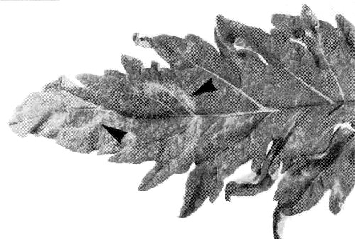
Naturally infected artichoke leaf (cv. Bayrampasa) showing faint chlorotic banding of the veins (arrow heads).

Chlorotic local lesions in Chenopodium quinoa.

Mottling and crinkling in a trifoliolate leaf of a systemically infected French bean plant.

Schlieren diagram of a purified virus preparation after centrifuging at 32 000 rev/min, showing three virus-specific components (T, M and B).

Virus particles in neutral 2% potassium phosphotungstate. A few empty shells (T component) are penetrated by the stain. Bar represents 100 nm.

Discrete accumulations of membranous vesicles (Ve) and virus particles (V) in the cytoplasm of a systemically infected C. quinoa cell. Bar represents 100 nm.
References list for DPV: Artichoke vein banding virus (285)
- Bishop, Claybrook & Spiegelman, J. molec. Biol. 26: 373, 1967.
- Chu & Francki, Virology 93: 398, 1979.
- Dunn & Hitchborn, Virology 25: 171, 1965.
- Gallitelli, Rana & Di Franco, Phytopath. Medit. 17: 1, 1978.
- Laemmli, Nature, Lond. 277: 680, 1970.
- Martelli, Rana & Savino, CMI/AAB Descr. of Pl. Viruses 176, 4 pp., 1977.
- Martelli, Quacquarelli, Gallitelli, Savino & Piazzolla, Phytopath. Medit. 17: 145, 1978.
- Martelli, Russo & Rana, Atti 3° Congr. Internaz. Carciofo, Bari, 1979: 895, 1981.
- Murant, in Handbook of Plant Virus Infections and Comparative Diagnosis, ed. E. Kurstak, p. 198, Elsevier/North Holland Biomedical Press, Amsterdam, 943 pp., 1981.
- Murant & Taylor, J. gen. Virol. 41: 53, 1978.
- Rana, Russo, Gallitelli & Martelli, Ann. appl. Biol. 101: 279, 1982.
- Reichmann, Virology 25: 166, 1965.
- Savino, Gallitelli & Barba, Ann. appl. Biol. 103: 243, 1983.
- Stace-Smith & Hansen, CMI/AAB Descr. Pl. Viruses 159, 4 pp., 1976.