Details of DPV and References
DPV NO: 302 September 1985
Family: Tombusviridae
Genus: Carmovirus
Species: Melon necrotic spot virus | Acronym: MNSV
Melon necrotic spot virus
T. Hibi Laboratory of Applied Microbiology, Department of Applied Physiology, National Institute of Agrobiological Resources, Tsukuba Science City, Yatabe, Ibaraki 305, Japan
I. Furuki Shizuoka Agricultural Experiment Station, 678-1, Toyoda-cho, Iwata-gun, Shizuoka-ken 438, Japan
Contents
- Introduction
- Main Diseases
- Geographical Distribution
- Host Range and Symptomatology
- Strains
- Transmission by Vectors
- Transmission through Seed
- Transmission by Grafting
- Transmission by Dodder
- Serology
- Nucleic Acid Hybridization
- Relationships
- Stability in Sap
- Purification
- Properties of Particles
- Particle Structure
- Particle Composition
- Properties of Infective Nucleic Acid
- Molecular Structure
- Genome Properties
- Satellite
- Relations with Cells and Tissues
- Ecology and Control
- Notes
- Acknowledgements
- Figures
- References
Introduction
-
Described by
Kishi (1960).
A virus with RNA-containing isometric particles c. 30 nm in diameter. It is transmitted in soil by the chytrid fungus Olpidium radicale (= O. cucurbitacearum), and readily transmitted by mechanical inoculation to several species of Cucurbitaceae. The virus occurs naturally only in greenhouse melons and cucumbers, and causes significant decrease in yield. Found in Japan, USA, the Netherlands and UK.
Main Diseases
Causes necrotic spots (Fig. 1, Fig. 3) or large necrotic lesions (Fig. 2) on the leaves, and necrosis on the stems of greenhouse melons (Kishi, 1966; Furuki, 1981) and cucumbers (Fig. 4) (Bos et al., 1984). In Japan, the disease is prevalent in winter. In the Netherlands, infection of cucumber was especially associated with the use of rockwool as a growing medium (Bos et al., 1984).
Geographical Distribution
Found in Japan (Kishi, 1960), USA (Gonzalez-Garza et al., 1979), the Netherlands (Bos et al., 1984) and UK (J. A. Tomlinson & B. J. Thomas, personal communication).
Host Range and Symptomatology
Japanese isolates of the virus were transmitted experimentally by inoculation of sap to Cucumis melo, Cucumis sativus, Citrullus lanatus, Cucurbita moschata, Lagenaria siceraria and Vigna unguiculata ssp. sesquipedalis (Kishi, 1966; Furuki et al., 1980; Furuki, 1981), the virus becoming systemic only in Cucumis melo. Californian isolates of the virus were transmitted mechanically to 18 species of cucurbitaceous plants but not to Lagenaria siceraria or Vigna unguiculata ssp. sinensis cv. Blackeye; systemic infections were produced only in Cucumis melo and C. anguria var. longipes (Gonzalez-Garza et al., 1979). European isolates have a similar host range but induce systemic symptoms in cucumber and melon only erratically (Bos et al., 1984; J. A. Tomlinson & B. J. Thomas, personal communication).
-
Diagnostic species
- Cucumis melo
(muskmelon). Necrotic lesions develop in inoculated cotyledons (Fig. 3) in 3-5 days and enlarge to 2-3 mm or more. They coalesce and the cotyledons desiccate and die 15-21 days after inoculation. The upper leaves begin to show necrotic spots 7 days after inoculation. When the virus is transmitted by the vector fungus, the roots become brown because of the fungus infection and the virus induces necrosis of the basal parts of the stems, and necrotic spots on the leaves. Other systemic symptoms include large necrotic lesions on the leaves, petiole necrosis and a decrease in size of the fruits, which may show necrotic spots. The large necrotic lesions on systemically infected leaves are produced only in plants infected by mechanical inoculation and not in those infected by Olpidium (Furuki, 1975; 1981). - Cucumis sativus (cucumber). Japanese isolates induce yellow spots
1 mm in diameter in inoculated cotyledons in 3-5 days; the spots become white
and cease to enlarge and there is no systemic infection. European isolates
induce chlorotic or white necrotic lesions 1-2 mm or more in diameter in
inoculated cotyledons
(Fig 4, left) in 2-5 days depending on the
temperature. At 30°C, they continue to enlarge and coalesce causing
collapse of the cotyledons. Systemic symptoms (leaves with small yellow
spots with brown pin-point centres expanding into large brown irregular
lesions up to 5 mm in diameter with yellow halos:
Fig. 4, right)
are produced erratically in up to 60% of inoculated plants depending on
the temperature, cultivar, and whether inoculation was mechanical or by
means of Olpidium. Lesions on systemically infected leaves finally
coalesce and the leaves shrivel and die. Stem necrosis is generally absent,
but at high temperatures (>28°C), plants infected by viruliferous
Olpidium exhibit severe necrosis of the hypocotyl (J. A. Tomlinson
& B. J. Thomas, personal communication). The virus does not induce
symptoms in fruits, but when
cucumber green mottle mosaic virus
is also
present, the fruits develop sunken chlorotic spots with dark green water-soaked
edges
(Bos et al., 1984).
- Citrullus lanatus (watermelon). Dark brown local lesions are induced in leaves or cotyledons about 6 days after inoculation. No systemic infection.
- Vigna unguiculata ssp. sesquipedalis. Japanese isolates induce tiny necrotic spots in inoculated primary leaves in 2-3 days. No systemic infection.
- Citrullus lanatus (watermelon). Dark brown local lesions are induced in leaves or cotyledons about 6 days after inoculation. No systemic infection.
-
Propagation species
- Cucumis melo.
Highly infective inoculum is obtained from cotyledons 3 days after inoculation.Assay species
- Cucumis melo
and Cucumis sativus are suitable for local lesion assay. The former is suitable also for whole plant assay and for assaying transmission by the vector.
Strains
Isolates studied in Japan (Kishi, 1960), USA (California strain; Gonzalez-Garza et al., 1979) and Europe (Bos et al., 1984; J. A. Tomlinson & B. J. Thomas, personal communication) appear to differ slightly in host range and symptomatology (see above) but no direct comparisons have been made.
Transmission by Vectors
Transmitted in soil by zoospores of the chytrid fungus Olpidium radicale (= O. cucurbitacearum) (Barr, 1968; Lange & Insunza, 1977; Komuro et al., 1970; Furuki, 1981). Virus particles attach to the external surface of the zoospores but are not carried internally (Furuki, 1981). The fungus is observed abundantly only in root tissues of virus-infected plants (Fig. 5). O. radicale can be freed of virus by air-drying the infected roots or by culturing the fungus on Benincasa hispida (Furuki, 1981). O. radicale differs in morphology and host range from O. brassicae. Japanese and Canadian isolates of O. radicale are similar in being able to transmit cucumber necrosis virus (Barr, 1968; Dias, 1970; Furuki et al., 1980; Furuki, 1981), but they differ in ability to parasitize squash and cucumber, and in the morphology of internal structures in the resting spores. Japanese isolates are therefore known as the melon strain of O. radicale (Furuki, 1981). Melon necrotic spot virus was not transmitted by Aphis gossypii (Kishi, 1966).
Transmission through Seed
Seed transmission of the virus in melon has been reported (Kishi, 1966: 10-15%; Gonzalez-Garza et al., 1979: 1-6%). Furuki (1981) observed no infection when the affected muskmelon seeds were sown in soil without the fungus vector, but when they were sown in soil containing the virus-free fungus, 10-40% of the seedlings became infected. He proposed that this type of seed transmission be called ‘vector-mediated seed transmission’.
Serology
The virus is an excellent immunogen. Rabbit antisera with titres of 1/1280 in precipitin tests are readily obtained. The virus forms a single precipitin band in gel double-diffusion tests (Hibi et al., 1980; Furuki, 1981).
Relationships
The type isolate from Japan is closely serologically related to the California isolate (Gonzalez-Garza et al., 1979) and the European isolates (Bos et al., 1984; J. A. Tomlinson & B. J. Thomas, personal communication) but not to any of the following viruses: cucumber necrosis (Hibi et al., 1980; Furuki, 1981; Bos et al., 1984), tobacco necrosis (Bos et al., 1984; T. Hibi, unpublished data), cucumber mosaic, squash mosaic, wild cucumber mosaic, turnip yellow mosaic (Gonzalez-Garza et al., 1979), cucumber fruit streak, cucumber soil-borne, cucumber leaf spot, carnation mottle, carnation ringspot, pelargonium leaf curl and tomato bushy stunt (Bos et al., 1984). Melon necrotic spot virus resembles cucumber necrosis virus in many properties such as transmissibility by the same fungus, possession of isometric particles c. 30 nm in diameter and infectivity for cucurbitaceous plants, but they differ in their physical and chemical properties, host range and symptoms on cucumber and muskmelon as well as in being serologically unrelated (Hibi et al., 1980; Furuki et al., 1980; Furuki, 1981). They should probably be classified together in the same virus group, but no such group has been formally proposed.
Stability in Sap
In muskmelon sap, the thermal inactivation point (10 min) is 60°C, the dilution end-point is 10-4-10-5, and the virus retains infectivity for 9-32 days at room temperature and for 131 days at 4°C (Kishi, 1966; Gonzalez-Garza et al., 1979; Furuki, 1981).
Purification
(Saito & Kishi, 1967; Hibi et al., 1980; Furuki, 1981). Inoculated cotyledons of muskmelon are picked 3 days after inoculation and extracted directly. Freezing the cotyledons greatly reduces the final yield of purified virus. All steps should be performed at 4°C. Grind 100 g cotyledons with 200 ml 0.067 M sodium phosphate buffer, pH 7.2, and 50 ml 0.1 M ascorbic acid. Filter the sap through gauze and centrifuge at 15,000 g for 10 min. Add 0.1 vol. chloroform to the supernatant fluid, acidify it to pH 5.3 with 1 M HCl, stir for 10 min and centrifuge at 15,000 g for 10 min. Centrifuge the supernatant fluid at 105,000 g for 120 min and resuspend the pellets in 0.01 M sodium phosphate buffer, pH 7.0. Follow with 2 or 3 further cycles of differential centrifugation. The preparation can be further purified by centrifugation in 10-40% linear sucrose density gradients at 58,000 g for 150 min. The yield of virus is 4-7 mg per 100 g fresh tissue.
Properties of Particles
The virus particles sediment as a single component in sucrose density gradients (Hibi et al., 1980; Furuki, 1981; Bos et al., 1984).
Sedimentation coefficient, (s20, w): 134 S at infinite dilution (Bos et al., 1984).
Buoyant density in caesium chloride: 1.33-1.34 g/cm3 (Hibi et al., 1980; Furuki, 1981; Bos et al., 1984).
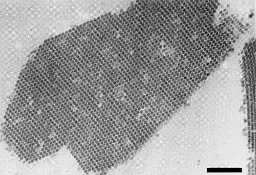
Particle Structure
Isometric particles c. 30 nm in diameter (Fig. 6) (Saito et al., 1962; Saito & Kishi, 1967; Gonzalez-Garza et al., 1979; Hibi et al., 1980; Furuki, 1981; Bos et al., 1984).
Particle Composition
Nucleic acid: RNA, single-stranded, about 17.8% of the particle weight (Hibi et al., 1980; Furuki, 1981). One species, of M. Wt 1.5 x 10⁶, determined under non-denaturing conditions in 2.5% polyacrylamide gels (Hibi et al., 1980; Furuki, 1981).
Protein: one species of polypeptide, M. Wt 46,000 (Bos et al., 1984).
Relations with Cells and Tissues
In diseased muskmelon the virus is present in roots, stems, petioles, leaves, fruits and seeds (Furuki, 1981). The virus is detected in the internal part of the seed coat but not in the embryo (Furuki, 1981). The vector fungus is observed only in roots (Furuki, 1981). Virus particles are found in aggregates or crystalline arrays in the cytoplasm or in the central vacuole of infected plant cells (Fig. 7). In roots, virus particles are often seen to be attached to the surface of the fungus but not within the fungal body (Hibi, 1983).
Notes
Melon necrotic spot virus is distinguished from cucumber necrosis and tobacco necrosis viruses by its host range and serological properties. As mentioned above, melon necrotic spot virus has a very narrow host range, while cucumber necrosis and tobacco necrosis viruses have wider host ranges. Melon necrotic spot virus has no serological relationship to cucumber necrosis or tobacco necrosis viruses. Control of the Olpidium vector can be obtained by soil sterilization with steam or methyl bromide (Komuro et al., 1970; Furuki, 1975; Furuki, 1981; Bos et al., 1984). For crops grown on rockwool, it is effective to add a surfactant (alkyl phenol ethylene oxide, Agral) to the nutrient solution at 20 µg/ml (J. A. Tomlinson & B. J. Thomas, personal communication).
Figures
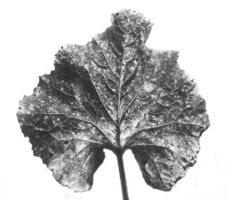
Necrotic spots in systemically infected muskmelon leaf.

Large necrotic lesions in systemically infected muskmelon leaf.

Necrotic lesions in inoculated muskmelon cotyledons.

Local lesions in inoculated cotyledons (left) and chlorotic spots in systemically infected leaf (right) of cucumber. (Courtesy of Dr. L. Bos, Research Institute for Plant Protection, Wageningen, The Netherlands.)

Zoosporangia of Olpidium radicale in infected muskmelon root.

Purified virus particles. Bar represents 100 nm.
Crystalline aggregate of virus particles in infected root cell of muskmelon. Bar represents 250 nm.
References list for DPV: Melon necrotic spot virus (302)
- Barr, Can. J. Bot. 46: 1087, 1968.
- Bos, Van Dorst, Huttinga & Maat, Neth. J. Pl. Path. 90: 55, 1984.
- Dias, Virology 42: 204, 1970.
- Furuki, Shokubutsu boeki 29: 447, 1975.
- Furuki, Tech. Bull. Shizuoka agric. Exp. Stn 14: 94 pp., 1981.
- Furuki, Hibi, Honda, Saito & Komuro, Ann. phytopath. Soc. Japan 46: 419, 1980.
- Gonzalez-Garza, Gumpf, Kishaba & Bohn, Phytopathology 69: 340, 1979.
- Hibi, in Handbook of Plant Viruses in Japan, p. 368, eds K. Yora, Y. Saito, Y. Doi, T. Inouye & K. Tomaru, Asakura Shoten, Tokyo, 627 pp., 1983.
- Hibi, Furuki, Honda, Saito & Komuro, Ann. phytopath. Soc. Japan 46: 419, 1980.
- Kishi, Ann. phytopath. Soc. Japan 25: 237, 1960.
- Kishi, Ann. phytopath. Soc. Japan 32: 138, 1966.
- Komuro, Furuki & Ezuka, Shokubutsu boeki 24: 399, 1970.
- Lange & Insunza, Trans. Br. mycol. Soc. 69: 377, 1977.
- Saito & Kishi, Ann. phytopath. Soc. Japan 33: 59, 1967.
- Saito, Kishi & Iwata, Ann. phytopath. Soc. Japan 27: 270, 1962.