Details of DPV and References
DPV NO: 304 September 1985
Family: Alphaflexiviridae
Genus: Potexvirus
Species: Wineberry latent virus | Acronym: WLV
Wineberry latent virus
A. T. Jones Scottish Crop Research Institute, Invergowrie, Dundee, DD2 5DA, UK
Contents
- Introduction
- Main Diseases
- Geographical Distribution
- Host Range and Symptomatology
- Strains
- Transmission by Vectors
- Transmission through Seed
- Transmission by Grafting
- Transmission by Dodder
- Serology
- Nucleic Acid Hybridization
- Relationships
- Stability in Sap
- Purification
- Properties of Particles
- Particle Structure
- Particle Composition
- Properties of Infective Nucleic Acid
- Molecular Structure
- Genome Properties
- Satellite
- Relations with Cells and Tissues
- Ecology and Control
- Notes
- Acknowledgements
- Figures
- References
Introduction
Described by Jones (1977).
A virus with flexuous filamentous particles c. 510 x 12 nm. Transmissible by mechanical inoculation of sap to several herbaceous species. Reported only from Scotland in a single symptomless plant of wineberry (Rubus phoenicolasius). Shows many properties of potexviruses and is serologically related to two such viruses - cactus X and viola mottle.
Main Diseases
Occurred symptomlessly in its natural host, wineberry (Rubus phoenicolasius), which was co-infected with raspberry bushy dwarf virus. The virus complex induced no symptoms in other Rubus spp. or cvs that were infected by graft inoculation, except that the raspberry bushy dwarf virus component induced a faint and transient line-pattern leaf symptom in some R. phoenicolasius and R. procerus plants 3-4 wk after grafting (Jones, 1977).
Geographical Distribution
Reported only from Scotland in a single plant that had been imported from the USA but grown in the field in Scotland for at least 10 years (Jones, 1977). The geographical origin of the virus is therefore not known.
Host Range and Symptomatology
Naturally infected Rubus phoenicolasius and experimentally infected Rubus species and cvs show no foliar symptoms. Species in five families of herbaceous plants were infected experimentally but slow systemic infection occurred only in some Chenopodiaceae. None of five Nicotiana spp. tested were infected.
Diagnostic species
- Chenopodium album, C. amaranticolor, C. murale, C. quinoa.
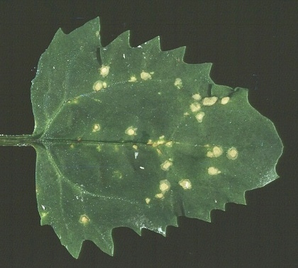
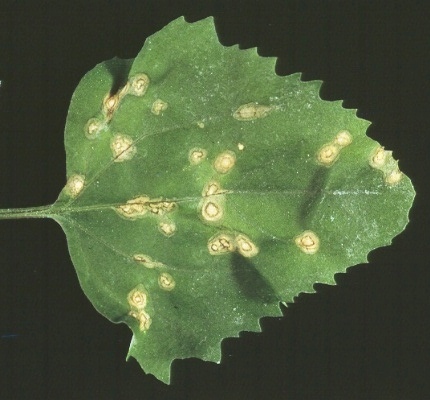
Small necrotic local lesions after 5-8 days (Fig. 1) which enlarge to form necrotic spots or rings (Fig. 2). Systemic infection, which is symptomless, occurs only in C. album and C. quinoa.- C. ambrosioides and Gomphrena globosa. Local red rings in 7 days
(Fig. 3). Symptomless
systemic infection in C. ambrosioides.
Propagation species
- Chenopodium amaranticolor
and C. quinoa (Jones, 1977). Assay species
- Chenopodium amaranticolor
is a good local lesion host.
Strains
None reported.
Transmission by Vectors
No vector reported. Macrosiphum euphorbiae given acquisition access feeds of 5 min, 1 h, 5 h or 24 h failed to transmit the virus in tests in which C. quinoa was used as the source and test plant (A. T. Jones, unpublished data).
Transmission through Seed
Not seed-borne in wineberry (Jones, 1977).
Serology
The virus is poorly immunogenic giving an antiserum with a titre of 1/128 in microprecipitin tests (Jones, 1977). Indirect ELISA with F(ab')2, fragments of antibodies to wineberry latent virus successfully detected the virus in sap from herbaceous plants and in fractions from sucrose density gradients (A. T. Jones & M. J. Mitchell, unpublished data).
Relationships
In particle size and morphology the virus resembles potexviruses. Purified virus preparations reacted strongly in indirect ELISA with antiserum to the potexviruses cactus X and viola mottle but not with antiserum to hydrangea ringspot, narcissus mosaic, nerine X, plantain X, potato X, tulip X or white clover mosaic viruses. However, in immunoelectron microscopy, wineberry latent virus particles were coated with antibodies to viola mottle virus but not to cactus virus X (A. T. Jones, M. J. Mitchell & I. M. Roberts, unpublished data). Both in microprecipitin tests and in immunosorbent electron microscopy, purified virus preparations failed to react with antiserum to apple chlorotic leaf spot, apple stem grooving, hydrangea ringspot, narcissus mosaic, potato X, potato T, raspberry bushy dwarf or white clover mosaic viruses (Jones, 1977; A. T. Jones & I. M. Roberts, unpublished data).
Stability in Sap
Sap of infected Chenopodium quinoa was infective when diluted 10-3 but not 10-4, heated for 10 min at 65 but not 70°C or stored at 18°C for 4 but not 8 days, at 4°C for 16 but not 32 days or at -15°C for 16 but not 32 days (Jones, 1977).
Purification
The virus particles are difficult to purify in quantity and in an unaggregated state. The best method found is to extract inoculated leaves of C. amaranticolor or C. quinoa 10-14 days after inoculation in 0.05 M Tris-HCl buffer (pH 7) containing 0.2% thioglycerol and 10% (v/v) chloroform (1 g leaf : 2 ml buffer : 0.2 ml chloroform) in a blender. Precipitate the virus by adding polyethylene glycol (PEG, M. Wt 6000) to 7% (w/v) and NaCl to 0.1 M, then sediment it through a 30% (w/v) sucrose pad containing 7% PEG + 0.1 M NaCl. Further purify by sucrose density gradient centrifugation (Jones, 1977).
Properties of Particles
Purified preparations usually consist of end-to-end aggregates of particles that sediment in a polydisperse manner with a sedimentation coefficient (s20,w) of 115-125 S (Jones, 1977).
A260/A280: 1.26 (Jones, 1977).
Buoyant density in Cs2SO4: 1.24 g/cm3 (A. T. Jones, unpublished data).
Particle Structure
Particles are flexuous filaments (Fig. 4) 12 nm wide and with a modal length in C. ambrosioides sap of c. 510 nm. The particles stain well in 2% solutions of ammonium molybdate, sodium phosphotungstate, uranyl acetate or uranyl formate/NaOH but tend to break on the grid (Jones, 1977) (Fig. 4). When measured by optical diffraction, the pitch of the helix appeared to be c. 348 Å with 8.8 subunits per turn (P. Tollin, personal communication).
Particle Composition
Nucleic acid: In agarose gels, virus RNA denatured with glyoxal migrated as a single component with an estimated M. Wt of c. 2.7 x 10⁶ (A. T. Jones & M. J. Mitchell, unpublished data).
Protein: In polyacrylamide/SDS gels, virus protein preparations contained a major polypeptide of estimated M. Wt 31,000 and a minor polypeptide of M. Wt 28,000 (A. T. Jones & M. J. Mitchell, unpublished data).
Relations with Cells and Tissues
No information.
Notes
The virus reacted with antiserum to the potexviruses cactus X and viola mottle in indirect ELISA but it differs greatly from viola mottle virus in experimental host range and in vitro properties (Lisa, Boccardo & Milne, 1982). It resembles cactus virus X in host range and symptomatology, but its particles were not coated with antibodies to this virus in immunoelectron microscopy tests. Furthermore, unlike cactus virus X, wineberry latent virus failed to react in microprecipitin and immunoelectron microscopy tests with antiserum to the potexviruses hydrangea ringspot, potato X and white clover mosaic (Bercks, 1971; Koenig & Lesemann, 1978). The M. Wt of the coat protein of wineberry latent virus also appears to be much larger than those of these and most other potexviruses (Koenig & Lesemann, 1978).
The virus is distinguished by particle length from the two other viruses with filamentous particles that occur in Rubus: the potyvirus bean yellow mosaic reported from red raspberry by Provvidenti & Granett (1974), which has particles 750 nm long, and bramble yellow mosaic virus reported from R. rigidus by Engelbrecht & van der Walt (1974), which has particles 730 nm long (Engelbrecht, 1976). Bramble yellow mosaic virus also differs from wineberry latent virus in infecting Rubus henryi and Nicotiana tabacum (Engelbrecht & van der Walt, 1974). A report (Cadman, 1963), that a third filamentous virus, apple chlorotic leaf spot, was present in raspberry plants with bushy dwarf disease is now disproved (Barnett & Murant, 1970; Murant, 1976) and antiserum to this virus did not react with particle preparations of wineberry latent virus (Jones, 1977).
Figures
References list for DPV: Wineberry latent virus (304)
- Barnett & Murant, Ann. appl. Biol. 65: 435, 1970.
- Bercks, CMI/AAB Descr. Pl. Viruses 58, 4 pp., 1971.
- Cadman, Pl. Dis. Reptr 47: 459, 1963.
- Engelbrecht, Acta Hort. 66: 79, 1976.
- Engelbrecht & van der Walt, Phytophylactica 6: 311, 1974.
- Jones, Ann. appl. Biol. 86: 199, 1977.
- Koenig & Lesemann, CMI/AAB Descr. Pl. Viruses 200, 4 pp., 1978.
- Lisa, Boccardo & Milne, CMI/AAB Descr. Pl. Viruses 247, 4 pp., 1982.
- Murant, CMI/AAB Descr. Pl. Viruses 165, 4 pp., 1976.
- Provvidenti & Granett, Pl. Dis. Reptr 58: 155, 1974.


