Details of DPV and References
DPV NO: 319 December 1986
Family: Tombusviridae
Genus: Aureusvirus
Species: Cucumber leaf spot virus | Acronym: CLSV
Cucumber leaf spot virus
Inge Weber Institut für Phytopathologie, Akademie der Landwirtschaftwissenschaften, Aschersleben, Germany
Contents
- Introduction
- Main Diseases
- Geographical Distribution
- Host Range and Symptomatology
- Strains
- Transmission by Vectors
- Transmission through Seed
- Transmission by Grafting
- Transmission by Dodder
- Serology
- Nucleic Acid Hybridization
- Relationships
- Stability in Sap
- Purification
- Properties of Particles
- Particle Structure
- Particle Composition
- Properties of Infective Nucleic Acid
- Molecular Structure
- Genome Properties
- Satellite
- Relations with Cells and Tissues
- Ecology and Control
- Notes
- Acknowledgements
- Figures
- References
Introduction
-
Described by
Weber et al. (1982).
Synonym
- Cucumber fruit streak virus (Gallitelli et al., 1983) is a closely related strain
-
A virus with RNA-containing isometric particles sedimenting as a single component. Readily transmitted by inoculation with sap; seed-borne and soil-borne. Found naturally only in cucumber. Reported from Europe and Jordan.
Main Diseases
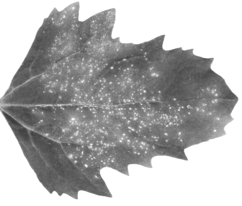
Young leaves of infected cucumbers (Cucumis sativus) show light green to yellowish, irregularly shaped spot-like clearings with brown necrotic centres (Fig. 1). Affected plants are severely stunted (Weber et al., 1982).
Geographical Distribution
Germany, Greece, Great Britain and Jordan (Weber et al., 1982, 1986; Gallitelli et al., 1983).
Host Range and Symptomatology
The cucumber leaf spot isolate infected all tested cultivars of Cucumis sativus and a wide range of herbaceous hosts (16 plant species in five dicotyledonous families), most of which were infected only locally (Weber et al., 1982). The cucumber fruit streak isolate infected 26 of 46 plant species in seven families (Gallitelli et al., 1983).
-
Diagnostic species
- Celosia argentea.
Red-brown necrotic spots 5-7 days after mechanical inoculation. No systemic infection. - Chenopodium quinoa. Local necrotic spots 5-7 days after mechanical
inoculation
(Fig. 2).
No systemic reaction.
- Cucumis sativus. Local necrotic spots on the cotyledons 7-9 days after mechanical inoculation (Fig. 3); systemic necrosis may occur, but not usually.
-
Propagation species
- Cucumis sativus
and Nicotania megalosiphon are good sources of virus for purification.Assay species
- Chenopodium quinoa
is a satisfactory local lesion host.
Strains
Weber et al. (1986) found a close serological relationship between cucumber leaf spot virus and cucumber fruit streak virus (Gallitelli et al., 1983): the serological differentiation index (SDI), determined from antiserum titres in agar gel double-diffusion tests, was <1. Nevertheless, the two isolates formed a spur in double-diffusion tests. Cucumber fruit streak virus is therefore regarded as a strain of cucumber leaf spot virus (Weber et al., 1986).
Transmission by Vectors
No vector known. Myzus persicae did not transmit the virus (Weber et al., 1982).
Transmission through Seed
Transmitted to seedlings through c. 1% of cucumber seed (I. Weber, unpublished data).
Serology
The virus is strongly immunogenic in rabbits: antisera with titres of 1/1024 are readily obtainable. Any of several serological tests can be easily performed. In gel-diffusion tests a single precipitin band is formed.
Relationships
There are numerous plant viruses with c. 30 nm isometric particles that sediment as a single component and contain a single RNA species of M. Wt c. 1.6 x 10⁶ and a single coat protein of M. Wt c. 44,000. In addition to the definitive tombusviruses listed by Matthews (1982), these viruses include carnation mottle, galinsoga mosaic, narcissus tip necrosis, pelargonium flower break, saguaro cactus, tephrosia symptomless and turnip crinkle viruses.
Cucumber leaf spot virus (type strain) did not react with antisera to the following isometric single-component viruses: carnation mottle, cocksfoot mild mosaic, cucumber necrosis, melon necrotic spot, narcissus tip necrosis, tomato bushy stunt and tobacco necrosis (Weber et al., 1982). The cucumber fruit streak strain did not react with antisera to the following viruses: artichoke mottled crinkle, cymbidium ringspot, carnation mottle, cucumber necrosis, pelargonium leaf curl, southern bean mosaic, sowbane mosaic, tobacco necrosis and tomato bushy stunt (Gallitelli et al., 1983). The affinities of cucumber leaf spot virus are therefore in doubt.
Stability in Sap
In crude sap of C. sativus, infectivity is lost after dilution between 10-6 and 10-7, heating for 10 min at between 80°C and 85°C or storing for 20 days at 22°C (Weber et al., 1982).
Purification
Weber et al. (1982) used a modification of Steere’s chloroform/butanol method. Homogenize 100 g inoculated cotyledons of C. sativus, 7-9 days after inoculation, in 200 ml 0.03 M phosphate buffer, pH 5. Emulsify the expressed sap with a mixture (1:1) of chloroform and n-butanol and clarify by low speed centrifugation. Concentrate the virus by two cycles of differential centrifugation, resuspending the pellets from high speed centrifugation in the same buffer. Further purify by rate-zonal centrifugation in sucrose gradients and resuspend in 0.03 M phosphate buffer, pH 5. About 6 mg virus may be obtained from 1 kg leaf material.
Gallitelli et al. (1983) used the following method to purify the cucumber fruit streak strain. Collect leaves of Nicotiana megalosiphon or C. sativus 5-7 days after inoculation and homogenize in 0.1 M phosphate buffer, pH 7.2, containing 0.1% thioglycollic acid. Filter the slurry through cheesecloth and clarify by slow addition, while stirring, of a 6% Na-bentonite suspension. Concentrate by two cycles of differential centrifugation (12,000 g for 15 min and 78,000 g for 2.5 h), resuspending the pellets from the high-speed centrifugations in 0.02 M phosphate buffer, pH 7.2. Purify further by centrifugation for 2.5 h at 24,000 rev/min through 10-40% sucrose density gradients in 0.02 M phosphate buffer, pH 7.2.
Properties of Particles
The particles form a single sedimenting and buoyant density component. The following data are from Weber et al. (1982) (leaf spot strain, LS) and Gallitelli et al. (1983) (fruit streak strain, FS).
Sedimentation coefficient (s20, w): 127 ± 2.5 S (LS); 132 S (FS).
Buoyant density in CsCl: 1.342 g/cm3 (LS); 1.35 g/cm3 (FS).
A260/A280: 1.60 (LS).
A(0.1%; 260, 1 cm) 5.63 (LS).
Particle Structure
Particles are isometric, about 28 nm in diameter (Fig. 4).
Particle Composition
Nucleic acid: RNA, single-stranded, 20% by weight of the particle; one electrophoretic species of M. Wt about 1.65 x 10⁶, estimated by gel electrophoresis under non-denaturing conditions (Weber et al., 1982). For the cucumber fruit streak strain Gallitelli et al. (1983) calculated 18% RNA content and a M. Wt about 1.45 x 10⁶.
Protein: Polyacrylamide gel electrophoresis of SDS-treated virus shows one polypeptide of M. Wt c. 44,000 for the type strain (Weber et al., 1982) and c. 47,000 for the fruit streak strain (Gallitelli et al., 1983).
Other components: None reported.
Relations with Cells and Tissues
Virus particles are easily detected in necrotic areas of inoculated leaves of C. sativus and C. quinoa (Fig. 5). In systemically infected cucumber leaves the particles are randomly scattered in the cytoplasm or form small clusters. Most of the cell organelles are apparently unaffected but sometimes mitochondria adopt an unusual shape (Fig. 6) and the Golgi apparatus proliferates, forming many small vesicles containing fine fibrils (Gallitelli et al., 1983; A. Stanarius & I. Weber, unpublished data).
Notes
In addition to the type and fruit streak strains of cucumber leaf spot virus, two other soil-borne viruses with c. 30 nm isometric particles are newly described from cucumber: melon necrotic spot virus (Gonzales-Garza et al., 1979) and cucumber soil-borne virus (Koenig et al., 1982). These two viruses are serologically unrelated to cucumber leaf spot virus (Weber & Stanarius, 1984).
Figures

Leaf spots in naturally infected leaf of Cucumis sativus.
Local necrotic spots in inoculated leaf of Chenopodium quinoa.

Local necrotic spots in inoculated cotyledons of Cucumis sativus.
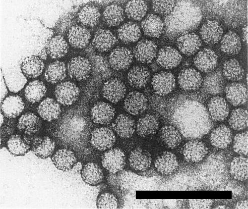
Particles from a purified preparation in 2% uranyl acetate. Bar represents 100 nm.

Virus particles in a necrotic cell of C. quinoa. Bar represents 500 nm. (Original electron micrograph by A. Stanarius.)

Abnormal mitochondria (Mi) in an infected cell of C. sativus (CW, cell wall; Va, vacuole). Bar represents 500 nm. (Original electron micrograph by A. Stanarius.)
References list for DPV: Cucumber leaf spot virus (319)
- Gallitelli, Vovlas & Avgelis, Phytopath. Z. 106: 149, 1983.
- Gonzales-Garza, Gumpf, Kishaba & Bohn, Phytopathology 69: 340, 1979.
- Koenig, Lesemann, Huth & Makkouk, Phytopathology 72: 964, 1982.
- Matthews, Intervirology 17: 1, 1982.
- Weber, Proll, Osterman, Leiser, Stanarius & Kegler, Arch. Phytopath. PflSchutz 18: 137, 1982.
- Weber & Stanarius, Arch. Phytopath. PflSchutz 20: 447, 1984.
- Weber, Stanarius & Kalinina, Arch. Phytopath. PflSchutz 22: 169, 1986.