Details of DPV and References
DPV NO: 333 September 1988
Family: Secoviridae
Genus: Unassigned Secoviridae
Species: Black raspberry necrosis virus | Acronym: BRNV
Black raspberry necrosis virus
A. T. Jones Scottish Crop Research Institute, Invergowrie, Dundee DD2 5DA, UK
Contents
- Introduction
- Main Diseases
- Geographical Distribution
- Host Range and Symptomatology
- Strains
- Transmission by Vectors
- Transmission through Seed
- Transmission by Grafting
- Transmission by Dodder
- Serology
- Nucleic Acid Hybridization
- Relationships
- Stability in Sap
- Purification
- Properties of Particles
- Particle Structure
- Particle Composition
- Properties of Infective Nucleic Acid
- Molecular Structure
- Genome Properties
- Satellite
- Relations with Cells and Tissues
- Ecology and Control
- Notes
- Acknowledgements
- Figures
- References
Introduction
-
Described by
Stace-Smith (1955).
Synonym
- 52V virus (Jones & Murant, 1972)
-
A virus with isometric particles c. 28 nm in diameter that is common in Rubus species throughout the world. Transmitted by aphids in a semi-persistent manner and with difficulty by mechanical inoculation of Rubus sap to a few herbaceous plant species.
Main Diseases
In nature, the virus is restricted to Rubus species. It kills shoot tips and causes subsequent chlorotic mottling or mosaic in black raspberry (Rubus occidentalis) (Fig. 1). On its own in red raspberry (R. idaeus and R. idaeus var. strigosus) and hybrid Rubus berries it induces no leaf symptoms or mild chlorotic flecking, mottling or line-patterns (Fig. 3, Fig. 4; Stace-Smith, 1955; Jones & Jennings, 1980; Jones et al., 1984). When present with raspberry bushy dwarf virus (Murant, 1976) in the red raspberry cv. Lloyd George, it induces bushy dwarf disease (Jones, 1979). When present with rubus yellow net virus (Stace-Smith & Jones, 1978) in some North American red raspberry cultivars, it is reported to cause raspberry veinbanding mosaic disease (Stace-Smith, 1955; Jones, 1986).
Geographical Distribution
Worldwide, wherever Rubus species are grown, often as a result of the propagation of infected stock plants. The virus spreads naturally in Rubus only in the northern hemisphere where its aphid vectors are common.
Host Range and Symptomatology
Infects by grafting all Rubus species and cultivars tested (Jones & Jennings, 1980). Transmitted mechanically to herbaceous plants, but with difficulty, infecting only a few species in the Chenopodiaceae and Solanaceae.
-
Diagnostic species
- Chenopodium quinoa
and C. amaranticolor. Difficult to infect. Mechanically inoculated plants sometimes show chlorotic/necrotic local lesions 6-10 days after inoculation; infected plants usually show systemic chlorotic flecking and/or necrosis 3-7 days later (Fig. 5). -
Chenopodium murale. Necrotic local lesions 5-10 days after inoculation
(Fig. 6).
Usually no systemic infection.
- Rubus occidentalis (black raspberry) and R. henryi. About 5-7 days after aphid inoculation, or 1-3 months after graft inoculation, the shoot tip begins to curl downward and slowly dies. Leaves beneath the tip wilt and/or become necrotic (Fig. 1, Fig. 2). Following this ‘shock’ reaction, newly produced leaves show chlorotic mottling or mosaic.
- R. molaccanus. Graft-inoculated plants develop apical necrosis and epinasty 1-3 months after grafting. Young leaves show necrotic flecking and/or line-pattern symptoms.
- R. procerus. Graft-inoculated plants develop a severe chlorotic mottling of leaves 3-5 weeks after grafting but symptoms become less conspicuous in leaves produced later (Jones & Roberts, 1977; Jones & Jennings, 1980).
- Rubus occidentalis (black raspberry) and R. henryi. About 5-7 days after aphid inoculation, or 1-3 months after graft inoculation, the shoot tip begins to curl downward and slowly dies. Leaves beneath the tip wilt and/or become necrotic (Fig. 1, Fig. 2). Following this ‘shock’ reaction, newly produced leaves show chlorotic mottling or mosaic.
-
Propagation species
- Chenopodium quinoa
(Murant et al., 1976; Jones & Mitchell, 1986). Cultures are best maintained in Rubus hosts.Assay species
- Chenopodium murale, C. quinoa
(Murant et al., 1976; Jones & Mitchell, 1986).
Strains
Symptom variants in black raspberry have been reported (Converse, 1963; Jones & Mitchell, 1986) but only one of these has been identified serologically. Mellor & Stace-Smith (1980) reported an isolate of the virus which was more heat stable in raspberry than other isolates tested.
Transmission by Vectors
Transmitted in nature by the raspberry aphids Amphorophora agathonica in North America and A. idaei in Europe. Under experimental conditions, these vectors and Aulacorthum solani and Macrosiphum euphorbiae transmitted the virus to Chenopodium quinoa seedlings (Jones & Murant, 1972; Murant et al., 1976; Jones, 1976; Kurppa & Martin, 1986). Almost all studies on vector relations have been done in North America using A. agathonica. All instars of A. agathonica can transmit after minimum acquisition and inoculation access feeds of 15-30 min and 2 min respectively. Feeding aphids remain able to transmit for up to 4 h after acquisition of virus; starved aphids remain able to transmit for up to 4 days, depending on the temperature (Stace-Smith, 1955). Transmission therefore seems to be in a semi-persistent manner.
Transmission through Seed
Not seed-borne in red raspberry (Jones & Murant, 1972).
Serology
The virus is poorly immunogenic, giving only very low-titred antiserum when injected into rabbits (Murant et al., 1976; Jones & Mitchell, 1986). When injected in mixture with solanum nodiflorum mottle, an unrelated virus, the maximum titre to black raspberry necrosis virus was 1/128 (Jones & Mitchell, 1986). Indirect ELISA with F(ab')2 fragments of antibodies from this antiserum detected the virus in Rubus species and cultivars, and in herbaceous plants, but time of sampling in Rubus was critical for detection (Jones & Mitchell, 1986). Electron microscope grids coated with virus antiserum trapped particles from sap of infected C. quinoa and raspberry, although the number of particles seen was relatively small (Jones & Mitchell, 1986).
Relationships
In its vector relations, reactions in Rubus occidentalis and R. henryi, and response to thermotherapy, the virus resembles two other aphid-borne raspberry viruses, raspberry leaf mottle and raspberry leaf spot. However, black raspberry necrosis virus induces no symptoms in the red raspberry indicators of the other two viruses and, unlike these viruses, is transmissible by mechanical inoculation of sap to herbaceous plants (Jones, 1982). Furthermore, sap from raspberry plants infected with cultures of raspberry leaf mottle and raspberry leaf spot viruses failed to react in the indirect form of ELISA with antiserum to black raspberry necrosis virus (Jones & Mitchell, 1986), and raspberry plants infected with raspberry leaf mottle or raspberry leaf spot viruses remain infectible with black raspberry necrosis virus.
Stability in Sap
In assays on Chenopodium quinoa, sap of infected C. quinoa was infective when diluted 10-1 but usually not 10-2, heated for 10 min at 50°C but not 52°C, stored at 18°C for 6 h but not 24 h, or stored at 4°C or -15°C for more than 3 days (Jones & Murant, 1972). In a mixed culture with solanum nodiflorum mottle virus in sap of Nicotiana clevelandii, black raspberry necrosis virus retained infectivity for up to 8 days at 20°C, probably as a consequence of its higher particle concentration in the mixed culture (Jones & Mitchell, 1986).
Purification
The virus is present in only very low concentrations in infected raspberry and C. quinoa plants, and is difficult to maintain in culture in glasshouse-grown C. quinoa. The virus has been maintained in culture in C. quinoa by keeping inoculated plants in growth cabinets at 18°C and 8000 lux with a photoperiod of 8 h. Even when plants were maintained under these conditions only very small amounts of virus could be obtained from them (Murant et al., 1976). Small amounts of virus have been partially purified directly from raspberry (A. T. Jones, unpublished data). Virus yield was increased 1000-fold by growing the virus in Nicotiana clevelandii in mixed culture with solanum nodiflorum mottle virus (Jones & Mitchell, 1986). Virus is best purified by extracting infected leaves in 0.05 M citrate buffer, pH 6.0 (1 g leaf/2 ml buffer), clarifying the extract with diethyl ether, and subjecting the aqueous phase to differential centrifugation. Purify further by sucrose density gradient centrifugation in gradients made up in 0.005 M citrate buffer, pH 6 (Murant et al., 1976, Jones & Mitchell, 1986). The particles of the virus thus ‘purified’ are difficult to separate from those of solanum nodiflorum mottle virus but may be used to produce a bivalent antiserum from which the antibodies to solanum nodiflorum mottle virus can be removed by cross-absorption with homologous virus (Jones & Mitchell, 1986).
Properties of Particles
Purified particles are isometric, c. 28 nm in diameter and a large proportion are penetrated by negative stain (Fig. 7). In sucrose density gradients the virus sediments as two components, one of about 50 S (probably nucleic acid free) and another predominant one of c. 130 S which is infective (Murant et al., 1976; Jones & Mitchell, 1986).
Particle Composition
No information.
Relations with Cells and Tissues
The virus was inactivated in infected raspberry plants after exposure to 37°C for several weeks (Chambers, 1954; Stace-Smith & Mellor, 1957; Bolton & Turner, 1962; Converse, 1963). However, one isolate thought to be black raspberry necrosis virus survived heat treatment at 39-42°C for 2 months (Mellor & Stace-Smith, 1980).
In infected leaves both of Rubus and of Chenopodium quinoa, islands of dead cells occurred in vascular tissue and, as infection progressed, in the leaf blade. Cells adjacent to these dead areas showed cell wall outgrowths and enlarged plasmodesmata each containing a single row of virus-like isometric particles c. 22 nm in diameter (Fig. 8). Small groups of virus-like particles were often seen in the cytoplasm. Most cell organelles were unaltered but some chloroplasts were disrupted (Jones & Roberts, 1977).
Notes
Raspberry is commonly infected with a complex of several aphid-borne viruses whose vector relations, reactions in R. occidentalis and R. henryi, and response to heat treatment are similar (Jones, 1982). The viruses may be separated by transferring individual viruliferous aphids from plants infected with virus mixtures to a series of R. occidentalis (black raspberry) seedlings and permitting the aphids to feed for only a short time on each seedling (Stace-Smith, 1956). However, this is difficult in Europe because R. occidentalis is resistant to the indigenous aphid species, Amphorophora idaei (Jones, 1986). Moreover, because R. occidentalis responds similarly to this virus and to raspberry leaf mottle and raspberry leaf spot viruses, unequivocal detection of black raspberry necrosis virus can only be made serologically, by infectivity assays to Chenopodium quinoa or by graft inoculation to red raspberry indicators of raspberry leaf mottle and raspberry leaf spot viruses in which black raspberry necrosis virus induces no symptoms (Jones, 1982; Jones & Mitchell, 1986).
Figures

Epinasty and necrosis of shoot tip of graft-inoculated Rubus occidentalis.

Epinasty and necrosis of shoot tip of graft-inoculated R. henryi.
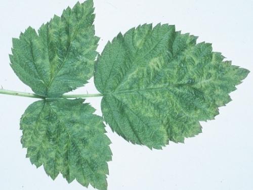
Chlorotic blotches and line-patterns in a leaf of graft-inoculated tayberry (R. ursinus x R. idaeus).

Veinal chlorotic spots in a leaf of graft-inoculated red raspberry (R. idaeus) cv. Orion.
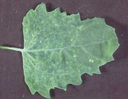
Systemic chlorotic flecks in Chenopodium quinoa leaf.

Necrotic local lesions in a leaf of C. murale, 7 days after mechanical inoculation.

Electron micrograph of partially purified virus particles stained with 1% uranyl acetate. Bar represents 100 nm.

Electron micrograph of a thin section of an infected R. occidentalis vascular cell showing virus-like particles aligned in a row within a tubule which passes through a plasmodesma. Bar represents 100 nm.
References list for DPV: Black raspberry necrosis virus (333)
- Bolton & Turner, Can. J. Pl. Sci. 42: 210, 1962.
- Chambers, Nature, Lond. 173: 595, 1954.
- Converse, Phytopathology 53: 1251, 1963.
- Jones, Ann. appl. Biol. 82: 503, 1976.
- Jones, J. hort. Sci. 54: 267, 1979.
- Jones, Acta Hort. 129: 41, 1982.
- Jones, Crop Res. 26: 127, 1986.
- Jones & Jennings, Ann. appl. Biol. 96: 59, 1980.
- Jones & Mitchell, Ann. appl. Biol. 109: 323, 1986.
- Jones & Murant, Pl. Path. 21: 166, 1972.
- Jones & Roberts, Ann. appl. Biol. 86: 381, 1977.
- Jones, Gordon & Jennings, J. hort. Sci. 59: 523, 1984.
- Kurppa & Martin, Acta Hort. 186: 51, 1986.
- Mellor & Stace-Smith, Acta Hort. 95: 71, 1980.
- Murant, CMI/AAB Descr. Pl. Viruses 165, 4 pp., 1976.
- Murant, Jones & Roberts, Acta Hort. 66: 39, 1976.
- Stace-Smith, Can. J. Bot. 33: 314, 1955.
- Stace-Smith, Can. J. Bot. 34: 435, 1956.
- Stace-Smith & Jones, CMI/AAB Descr. Pl. Viruses 188, 4 pp., 1978.
- Stace-Smith & Mellor, Can. J. Bot. 37: 287, 1957.