Details of DPV and References
DPV NO: 344 December 1989
Family: Virgaviridae
Genus: Hordeivirus
Species: Barley stripe mosaic virus | Acronym: BSMV
This is a revised version of DPV 68
Barley stripe mosaic virus
J. G. Atabekov Department of Virology, Moscow State University, 119899 Moscow, USSR
V. K. Novikov Department of Virology, Moscow State University, 119899 Moscow, USSR
Contents
- Introduction
- Main Diseases
- Geographical Distribution
- Host Range and Symptomatology
- Strains
- Transmission by Vectors
- Transmission through Seed
- Transmission by Grafting
- Transmission by Dodder
- Serology
- Nucleic Acid Hybridization
- Relationships
- Stability in Sap
- Purification
- Properties of Particles
- Particle Structure
- Particle Composition
- Properties of Infective Nucleic Acid
- Molecular Structure
- Genome Properties
- Satellite
- Relations with Cells and Tissues
- Ecology and Control
- Notes
- Acknowledgements
- Figures
- References
Introduction
-
Described by
McKinney (1951a,
1951b).
Synonym
- Barley false stripe virus (Rev. appl. Mycol. 30: 408)
-
A virus with straight tubular particles c. 22 nm in diameter and of two-four length (100-150 nm), depending on the strain. The particles contain separately encapsidated single-stranded RNA molecules of up to five lengths but comprising a functionally tripartite genome. The virus has a narrow host range and is transmitted through seed and pollen. Vector unknown. World-wide distribution.
Main Diseases
The only natural hosts known are barley (Hordeum vulgare) and wheat ( Triticum aestivum) in which the virus causes diseases ranging in severity from very mild stripe mosaic to lethal necrosis (McKinney & Greeley, 1965). Losses of 20% or more in barley yield have been reported (Timian & Sisler, 1955; Timian, 1974; Nutter et al., 1984). Wild oats (Avena fatua) can be infected occasionally (Chiko, 1975).
The virus induces mutations in host plants (Sprague et al., 1963; Sprague & McKinney, 1971), an effect designated ‘aberrant ratio’ because such mutations distort Mendelian ratios for several phenotypic characteristics (Brakke, 1984).
Geographical Distribution
World-wide.
Host Range and Symptomatology
Besides the natural host plants (barley, wheat, wild oats), about 240 members of the Gramineae, nine members of the Chenopodiaceae, and one member each of the Solanaceae, Amaranthaceae and Primulaceae have been infected experimentally (Jackson & Lane, 1981).
-
Diagnostic species
- Avena sativa
(oat), Hordeum vulgare (barley), and Triticum aestivum (wheat). Systemic stripe mosaics of different types (Fig. 1). -
Beta vulgaris (beet). Chlorotic yellow local lesions; not systemic.
- Chenopodium album, C. amaranticolor, C. quinoa. Large chlorotic yellow lesions (Fig. 3) ; not systemic.
- Some strains induce systemic stripe mosaic in Zea mays (maize; Fig. 2), mosaic in Spinacia oleracea (spinach) and local chlorotic lesions in Nicotiana tabacum cv. Samsun (tobacco).
- Chenopodium album, C. amaranticolor, C. quinoa. Large chlorotic yellow lesions (Fig. 3) ; not systemic.
-
Propagation species
- Hordeum vulgare
and Triticum aestivum are the best sources of virus for purification.Assay species
- Chenopodium amaranticolor
and C. quinoa are useful local lesion hosts.
Strains
Several variants of the virus (McKinney & Greeley, 1965), differing in the symptoms they induce in barley, wheat and oat have been named the latent, mild, moderate, severe, yellow leaf, white leaf, dwarf and necrotic strains. The mild and moderate strains are the most common. Most isolates have been maintained successfully in culture for many transfers but some variants become difficult or impossible to transfer by inoculation of sap (McKinney & Greeley, 1965). The reason for this change is not known. Seed transmissibility differs among strains: some strains seem rarely to infect the embryo or are inactivated during maturation of the seed (Singh et al., 1960; Hamilton, 1965; Carroll, 1972). The following strains are most commonly used: Type, Russian, Norwich, North Dakota 18 (ND18), Argentina mild (AM) and Rothamsted (Jackson & Brakke, 1973; Lane, 1974).
Transmission by Vectors
None reported.
Transmission through Seed
Transmitted through seed of barley to 90% (McKinney, 1951a, 1953) or even 100% (Gold et al., 1954) of seedlings. The virus is also pollen-borne and infects the pollinated plants (Gold et al., 1954; Gardner, 1967). No pollen or seed transmission occurs in maize (Sprague et al., 1963; Pring, 1974). The main route of natural spread of the virus seems to be by plant to plant contact. Nevertheless, serological testing of seed is advisable to ensure the use of virus-free seeds.
Serology
The virus is highly immunogenic. In immunodiffusion tests, intact virus particles usually form a single precipitin line close to the antigen well. Different strains of the virus are antigenically identical in immunodiffusion tests. Extracts of infected leaves contain virus particle protein in monomeric (fully dissociated) form which is serologically related but not identical to the intact virus particles (Hamilton & Boll, 1966); in addition there is a range of aggregates of the particle protein which have sedimentation coefficients greater than 10 S and are antigenically indistinguishable from each other and from intact virus particles (Atabekov et al., 1968a).
Relationships
The virus is morphologically similar and distantly serologically related to lychnis ringspot (Gibbs et al., 1963) and poa semilatent viruses (Slykhuis, 1972; Hunter et al., 1986) but is not related to tobamoviruses (Jackson & Lane, 1981), tobraviruses or to those furoviruses tested (Thouvenel & Fauquet, 1981).
Stability in Sap
In barley sap the thermal inactivation point (10 min) is about 65-68°C (McKinney, 1951b; Kassanis & Slykhuis, 1959). The virus can be stored in dehydrated tissue for as long as 12 years (McKinney & Greeley, 1965), in fresh leaf tissue in liquid nitrogen (McKinney et al., 1961) or in lyophilized sap (Timian & Klosterman, 1962). The virus retains infectivity for several years in infected seeds (Timian, 1965). Dilution end-point of sap varies greatly (from 5 x 10⁻² to 1 x 10⁻⁴) but is usually about 0.5-2.0 x 10⁻³. Longevity in vitro: not more than 1 month (McKinney, 1951b; Kassanis & Slykhuis, 1959).
Purification
Homogenize fresh or frozen systemically infected barley or wheat leaves in 2-3 vol 0.3 M glycine buffer, pH 8.0, with 1% polyethylene glycol (PEG), M.Wt 6000, 0.01 M MgSO4 and 0.2% mercaptoethanol. Filter the extract and adjust the pH to 7.5. Clarify the extract by centrifugation at 15,000 g for 20 min. Add Triton X-100 to the supernatant fluid to a concentration of 1% and incubate at 4°C for 30 min. Precipitate the virus by addition of PEG and NaCl to concentrations of 4% and 2%, respectively; incubate the mixture at 4°C for 1-2 h, then centrifuge at 10,000 g for 20 min. Resuspend the pellet in 0.3 M glycine buffer, pH 7.5, and centrifuge at 10,000 g for 10 min. Layer the supernatant fluid over 20% (w/v) sucrose solution comprising 1/6th of the centrifuge tube volume and centrifuge at 100,000 g for 2 h. Resuspend the pellet in 0.1 M glycine buffer, pH 7.5, containing 1 mM MgSO4 and clarify the preparation by centrifugation at 10,000 g for 10 min. Purify further by two cycles of differential centrifugation. Yields are 100-500 mg per kg fresh leaf material.
The specific infectivity of purified preparations varies considerably and is low compared with that of other viruses (Atabekov & Novikov, 1966). Purified preparations frequently have little infectivity (Brakke, 1959) or even none (Atabekov & Novikov, 1966). However, when virus preparations that are non-infective in C. amaranticolor are treated with phenol the extracted RNA is often found to be infective (Atabekov & Novikov, 1966; Novikov & Atabekov, 1970). The infectivity of the virus is optimal at pH 5.5-7.0. Factors in crude barley sap stabilize the virus infectivity (Brakke, 1962).
Properties of Particles
Virus preparations consist of rod-shaped particles (Fig. 4) of different lengths depending on the size of the RNA within them. The particles of ‘pseudobipartite’ strains form a single zone in sucrose gradients and upon analytical centrifugation (schlieren optics); those of the ‘pseudobipartite’ Russian strain have sedimentation coefficients (s°20,w) of about 185 S in 0.01 M phosphate buffer, pH 7.2 (Atabekov & Novikov, 1966). The intrinsic viscosity (h) of these preparations is 0.12-0.17 dl/g. In 0.1 M phosphate buffer the particles aggregate and this causes a change in the hydrodynamic properties of the preparations (s° 20,w = 263 S; h = 0.58 dl/g). The particles of the ‘pseudoquadripartite’ Argentina mild strain have sedimentation coefficients of about 194, 183, 171 and 166 S within a broad asymmetric peak in sucrose density gradients (Brakke & Palomar, 1976). The particles of ‘orthotripartite’ strain Norwich have sedimentation coefficients of 201, 197 and 182 S (Brakke & Palomar, 1976).
Partial specific volume: 0.732 ml g-1.
M. Wt of the long (185 S) particles of the Russian strain: c. 26 x 106 (Atabekov & Novikov, 1966).
Isoelectric point: c. pH 4.5.
Electrophoretic mobility: -21.0 x 105 cm2 sec-1 volt-1 at pH 7.7 in 0.05 M phosphate buffer (Russian strain).
A260(0.1%; 1 cm): 2.6, but a maximum value of 2.7 is obtained at 270 nm.
A260/A280: 1.00-1.05 (not corrected for light- scattering: Atabekov & Novikov, 1971; Brakke, 1979), 0.99 (corrected for light-scattering: Brakke, 1979), 0.96 (in SDS: Lane, 1974).
A270/A280 1.06-1.12 (not corrected for light-scattering).
Particle Structure
The particles are straight rods (about 22 nm in diameter) and helically constructed, the pitch of the basic helix being 2.5-2.6 nm (Finch, 1965; Atabekov & Novikov, 1966), with about 24 protein subunits per turn of the helix (Kiselev et al., 1966; Veerisetty, 1978). Diameter of the internal canal: 3.0-4.0 nm (Gibbs et al., 1963; Harrison et al., 1965; Finch, 1966). The RNA molecule is located at a radius of 5.5 nm (X-ray diffraction data; Finch, 1965).
From two to four main types of rod-like particle are present depending on the strain (Brakke & Palomar, 1976) in accordance with the sizes of the genomic RNA molecules (see Genome Properties). Separate encapsidation of the different RNA species results in particles of different lengths: 148, 128, 108 and 100 nm in ‘pseudoquadripartite’ strains; 143, 128 and 112 nm in ‘orthotripartite’ strains; 146 and 128 nm in ‘pseudobipartite’ strains (Harrison et al., 1965; Brakke & Palomar, 1976).
Particle Composition
Nucleic acid: Single-stranded RNA, comprising 3.8-4.0% (Atabekov & Novikov, 1971) or 3.7% (Brakke, 1979) of the particle weight. G:A:C:U = 20.3:30.9:19.4:29.4 (Atabekov & Novikov, 1971). Different strains have different sets of RNA species (Jackson & Brakke, 1973; Lane, 1974). Each RNA species is encapsidated separately; these species have been designated RNA 1 (or a), RNA2a (or ß), RNA2b (or g), RNA3 (or g) and RNA4 (or g). We now suggest that these RNAs should be designated RNA1, RNA2, RNA3II, RNA3III and RNA4, respectively. The natural tripartite (‘orthotripartite’) strains ND18 and Norwich contain RNA1 c. 3900 nucleotides long (M. Wt c. 1.35 x 10⁶), RNA2 c. 3300 nucleotides long (M. Wt c. 1.1 x 10⁶) and RNA3III c. 2800 nucleotides long (M. Wt c. 0.9 x 10⁶) (Jackson et al., 1983a; Gustafson et al., 1982, 1987; Gustafson & Armour, 1986; Afanasyev et al., 1986). In ‘pseudobipartite’ strains (Type, Russian) the second (RNA2) and third (RNA3II) are of similar size and form one band upon gel electrophoresis (Jackson & Brakke, 1973; Gustafson et al., 1982). The ‘pseudoquadripartite’ strain AM contains an additional species, RNA4 (Lane, 1974) c. 2600 nucleotides long (M. Wt c. 0.85 x 10⁶) (McFarland et al., 1983; Dolja et al., 1983a; Afanasyev et al., 1986). The sedimentation coefficient (s° 20,w) of RNA in 0.1 M NaCl is 20-21 S (Atabekov & Novikov, 1966; Pring, 1972). This value corresponds to RNA2 which is the major component of the mixture.
Protein: About 96% of the virus weight. The M. Wt of the protein subunit is c. 22-23 x 103 from hydrodynamic data and SDS/polyacrylamide gel electrophoresis (Kiselev et al., 1966; Atabekov et al., 1968b; Dolja et al., 1983a). The subunit contains 198 amino acid residues (predicted from the nucleotide sequence of the coat protein gene; Gustafson & Armour, 1986) including the N-terminal methionine which is absent from the natural capsid protein (Atabekov et al., 1968b; Dolja et al., 1982). The amino acid composition, determined by acid hydrolysis of the coat protein (Atabekov et al., 1968b; Gumpf & Hamilton, 1968) is in agreement with that predicted from the nucleotide sequence data of the coat protein gene (Gustafson & Armour, 1986). The protein contains relatively large amounts of polar amino acids and proline but no cysteine or methionine. Native subunits have little or no a-helix. Isoelectric point: pH 5.3. There is only one type of protein molecule in freshly isolated virus preparations. A partial proteolytic degradation of the coat protein takes place upon storage at 0°C if SDS is not present (Negruk et al., 1974). The virus dissociates to monomeric protein units (s°20,w = 1.8 S) under the influence of various agents including high concentrations of divalent cations, KCl, 1% SDS, acetic acid and alkali (Atabekov & Novikov, 1966, 1971; Kiselev et al., 1966; Atabekov et al., 1968b; Gumpf & Hamilton, 1968) The viral protein can be repolymerized in vitro into different types of aggregate including rod-like helical structures (Atabekov et al., 1968 a, 1968b, 1968c). The protein can be reassembled with RNA in vitro into rod-like nucleoprotein particles which are noninfective (Atabekov et al., 1970).
Other components: The capsid protein is a glycoprotein (Partridge et al., 1974), the carbohydrate moiety consisting of glucose, mannose, xylose, galactosamine and glucosamine, linked through an amide bond involving asparagine (Gumpf et al., 1977).
Genome Properties
The segmented genome of the virus (Jackson & Brakke, 1973; Lane, 1974) is functionally tripartite, but at the strain level the virus particles may contain up to five RNA segments. RNA1 has the same size in all strains and codes in vitro for a polypeptide of M. Wt 120,000 (p120) (Dolja et al., 1979, 1983b), presumably a component of the RNA replicase. RNA1 does not contain significant regions homologous to other RNA species (Palomar et al., 1977). The complete nucleotide sequence of RNA1 is given by Gustafson et al. (1989). RNA2 codes in vitro for a coat protein (p22-23) and under certain conditions for p25, a probable read-through product of the coat protein gene (Dolja et al., 1979, 1983b). The AUG codon of the coat protein gene is located at c. 90 nucleotides from the 5' end of RNA2 (Dolja et al., 1982). The complete nucleotide sequence of RNA2 (Gustafson & Armour, 1986) shows the presence of three additional open reading frames in RNA2 coding for three tentative proteins.
RNA3II, RNA3III and RNA4 code in vitro for polypeptides p85, p75 and p55, respectively (Dolja et al., 1979, 1983b; Gustafson et al., 1981). The amino acid sequences of p85, p75 and p55 overlap (Dolja et al., 1983b) and contain homologies with putative RNA polymerases of other viruses (Afanasyev et al., 1986; Gustafson et al., 1987). RNA3II and RNA3III carry in their 3'-terminal regions the gene for polypeptide p17 which is expressed from a subgenomic RNA which is also encapsidated (Dolja et al., 1983a; Jackson et al., 1983b).
The nucleotide sequences of RNA3III from the Argentina mild (Afanasyev et al., 1986) and ND18 (Gustafson et al., 1987) strains have been determined and compared with those of RNA3II. RNA3II of ‘pseudobipartite’ strains is homologous to RNA3III of ‘orthotripartite’ strains and appears to be its functional analogue (Gustafson et al., 1982; Dolja et al., 1983b). Its increased size is caused by an in-frame direct tandem repeat of nucleotides coding for about 120 amino acids (Afanasyev et al., 1986; Gustafson et al., 1987). The duplicated region in RNA3II increases the length of the polypeptide (p85) coded by this RNA as compared with the polypeptide (p75) coded by RNA3III. The nucleotide sequence of RNA4 of ‘pseudoquadripartite’ strain AM (Afanasyev et al., 1986) shows that it has sequence homology with RNA3III; its smaller size is accounted for by deletion from RNA3III of 185 nucleotides in the 3'-terminal part of the gene coding for p75 and by the formation of a new terminator UGA (Afanasyev et al., 1986). RNA4 appears to be a functionally inessential defective fragment of RNA3III. It can be irreversibly eliminated without loss of infectivity (Palomar & Brakke, 1976; McFarland et al., 1983). The virus has a metastable genome with a high degree of variability (Jackson & Lane, 1981; Atabekov & Dolja, 1986).
The virus has a unique arrangement at the 3'-terminal region of all the RNA species: they contain an internal poly(A) tract of varying length followed by a 3'-terminal tRNA-like structure that can accept tyrosine (Agranovsky et al., 1981, 1982, 1983; Kozlov et al., 1984). A 5'-terminal cap structure has been detected in RNA species 1 and 3 of the Norwich strain (Agranovsky et al., 1979).
Relations with Cells and Tissues
All tissues are infected. The virus particles are present in mesophyll, guard, epidermal, vascular parenchyma, sieve tube and tracheary cells (McMullen et al., 1978; Carroll, 1986). Mature particles accumulate in the cytoplasm and the nuclei; they also occur in the cytoplasm of the pollen cells (Shalla, 1959; Gardner, 1967). The virus can survive and probably multiply in haploid cells of barley (Gardner, 1967; Carroll, 1974). Particles of the type (seed-transmitted) strain, in contrast to those of the NSP (non-seed passage) strain, were found in embryos, ovules and pollen (Carroll, 1972). In floral cells the virus particles are in association with plastids and microtubules (Carroll & Mayhew, 1976a, 1976b; Carroll, 1986). The microtubules may be involved in the assembly and cell-to-cell transport of the virus (Mayhew & Carroll, 1974b). Crystalline inclusions have not been detected (Shalla, 1959; Mayhew & Carroll, 1974a). Globular inclusions not containing virus particles have been found in the cytoplasm (Gardner, 1967). The cytopathology of the virus in host tissues has been studied by McMullen et al. (1977a, 1977b) and Lin & Langenberg (1984, 1985a, 1985b).
Notes
In natural and experimental hosts the symptoms induced may vary depending on the virus strain and conditions of infection. The symptoms may be confused with those of other viruses, therefore the virus can be identified unequivocally only by serology.
Figures
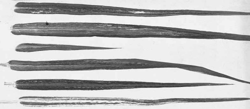
Different types of stripe mosaic induced in barley leaves.
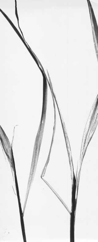
Mosaic induced in leaves of sweet corn plant.

Lesions in inoculated leaf of Chenopodium amaranticolor.

Electron micrograph of virus particles from purified preparation in 2% (w/v) uranyl acetate.
References list for DPV: Barley stripe mosaic virus (344)
- Afanasyev, Rupasov, Fedchenko, Dolja, Atabekov, Chernov, Kozlov & Bayev, Dokl. Akad. Nauk USSR, 290: 724, 1986.
- Agranovsky, Dolja, Kagramanova & Atabekov, Virology 95: 208, 1979.
- Agranovsky, Dolja, Gorbulev, Kozlov & Atabekov, Virology 113: 174, 1981.
- Agranovsky, Dolja & Atabekov, Virology 119: 51, 1982.
- Agranovsky, Dolja & Atabekov, Virology 129: 344, 1983.
- Atabekov & Dolja, In The Plant Viruses, Vol. 2, p. 397, ed. M.H.V. van Regenmortel & H. Fraenkel-Conrat, Plenum, New York, 424 pp., 1986.
- Atabekov & Novikov, Biokhimia, USSR 31: 157, 1966.
- Atabekov & Novikov, CMI/AAB Descr. of Pl. Viruses 68, 4 pp., 1971.
- Atabekov, Dementyeva, Schaskolskaya & Sacharovskaya, Virology 36: 601, 1968a.
- Atabekov, Novikov, Kiselev, Kaftanova & Egorov, Virology 36: 620, 1968b.
- Atabekov, Schaskolskaya, Dementyeva, Sacharovskaya & Senchenkov, Virology 36: 587, 1968c.
- Atabekov, Novikov, Vishnichenko & Kaftanova, Virology 41: 519, 1970.
- Brakke, Virology 9: 506, 1959.
- Brakke, Virology 17: 131, 1962.
- Brakke, Virology 98: 76, 1979.
- Brakke, A. Rev. Phytopath. 22: 77, 1984.
- Brakke & Palomar, Virology 71: 255, 1976.
- Carroll, Virology 48: 323, 1972.
- Carroll, Virology 60: 21, 1974.
- Carroll, In The Plant Viruses, Vol. 2, p. 373, ed. M.H.V. van Regenmortel & H. Fraenkel-Conrat, Plenum, New York, 424 pp., 1986.
- Carroll & Mayhew, Can. J. Bot. 54: 1604, 1976a.
- Carroll & Mayhew, Can. J. Bot. 54: 2497, 1976b.
- Chiko, Can. J. Bot. 53: 417, 1975.
- Dolja, Sokolova, Tjulkina & Atabekov, Mol. gen. Genet. 175: 93, 1979.
- Dolja, Lunina, Smirnov, Kaplan, Hudjakov, Kozlov, Bajev & Atabekov, Dokl. Akad. Nauk USSR 265: 474, 1982.
- Dolja, Agranovsky, Lunina & Atabekov, FEBS Lett. 151: 215, 1983a.
- Dolja, Lunina, Leiser, Stanarius, Belzhelarskaya, Kozlov & Atabekov, Virology 127: 1, 1983b.
- Finch, J. molec. Biol. 12: 612, 1965.
- Finch, Nature, Lond. 212: 349, 1966.
- Gardner, Phytopathology 57: 1315, 1967.
- Gibbs, Kassanis, Nixon & Woods, Virology 20: 194, 1963.
- Gold, Suneson, Houston & Oswald, Phytopathology 44: 115, 1954.
- Gumpf & Hamilton, Virology 35: 87, 1968.
- Gumpf, Cunningham, Heick & Shannon, Virology 78: 328, 1977.
- Gustafson & Armour, Nucleic Acids Res. 14: 3895, 1986.
- Gustafson, Larkins & Jackson, Virology 111: 579, 1981.
- Gustafson, Milner, McFarland, Pedersen, Larkins & Jackson, Virology 120: 182, 1982.
- Gustafson, Hunter, Harau, Armour & Jackson, Virology 158: 394, 1987.
- Gustafson, Armour, Gamboa, Burgett & Shepherd, Virology 170: 370, 1989.
- Hamilton, Phytopathology 55: 798, 1965.
- Hamilton & Boll, Virology 30: 661, 1966.
- Harrison, Nixon & Woods, Virology 26: 284, 1965.
- Hunter, Heaton, Bracker & Jackson, Phytopathology 76: 322, 1986.
- Jackson & Brakke, Virology 55: 483, 1973.
- Jackson & Lane, In Handbook of Plant Virus Infections and Comparative Diagnosis, p.565, ed. E.Kurstak, Elsevier/North-Holland, Amsterdam, 943 pp., 1981.
- Jackson, McFarland, Gustafson, Dawson & Stanley, In Plant Molecular Biology. UCLA Symp. Molec. Cell. Biol., Vol. 12, p. 67, ed. R. R. Goldberg. Alan R. Liss, New York, 1983a.
- Jackson, Dawson, Covey, Hull, Davies & McFarland, Virology 127: 37, 1983b.
- Kassanis & Slykhuis, Ann. appl. Biol. 47: 254, 1959.
- Kiselev, Atabekov, Kaftanova & Novikov, Biokhimia USSR 31: 670, 1966.
- Kozlov, Rupasov, Adashev, Belgelarskaya, Agranovsky, Mankin, Morozov, Dolja & Atabekov, Nucleic Acids Res. 12: 4001, 1984.
- Lane, Virology 58: 323, 1974.
- Lin & Langenberg, J. gen. Virol. 65: 2217, 1984.
- Lin & Langenberg, J. Ultrastruct. Res. 84: 16, 1985a.
- Lin & Langenberg, Virology 142: 291, 1985b.
- Mayhew & Carroll, Virology 58: 561, 1974a.
- Mayhew & Carroll, Science, N. Y. 185: 957, 1974b.
- McFarland, Brakke & Jackson, Virology 130: 397, 1983.
- McKinney, Pl. Dis. Reptr 35: 48, 1951a.
- McKinney, Phytopathology 41: 563, 1951b.
- McKinney, Pl. Dis. Reptr 37: 292, 1953.
- McKinney & Greeley, Tech. Bull. U.S. Dep. Agric. No. 1324, 84 pp., 1965.
- McKinney, Greeley & Clark, Pl. Dis. Reptr 45: 755, 1961.
- McMullen, Gardner & Myers, Proc. S. Dak. Acad. Sci. 56: 100, 1977a.
- McMullen, Gardner & Myers, Phytopathology 67: 462, 1977b.
- McMullen, Gardner & Myers, Proc. S. Dak. Acad. Sci. 57: 92, 1978.
- Negruk, Novikov & Atabekov, Dokl. Akad. Nauk USSR 218: 489, 1974.
- Novikov & Atabekov, Virology 41: 101, 1970.
- Nutter, Pederson & Timian, Phytopathology 74: 363, 1984.
- Palomar & Brakke, Phytopathology 66: 1422, 1976.
- Palomar, Brakke & Jackson, Virology 77: 471, 1977.
- Partridge, Shannon, Gumpf & Goldbaugh, Nature, Lond. 247: 391, 1974.
- Pring, Virology 48: 22, 1972.
- Pring, Phytopathology 64: 64, 1974.
- Shalla, Virology 7: 193, 1959.
- Singh, Arny & Pound, Phytopathology 50: 290, 1960.
- Slykhuis, Phytopathology 62: 508, 1972.
- Sprague & McKinney, Genetics 67: 533, 1971.
- Sprague, McKinney & Greeley, Science, N.Y. 141: 1052, 1963.
- Timian, Pl. Dis. Reptr 49: 696, 1965.
- Timian, Phytopathology 64: 342, 1974.
- Timian & Klosterman, Phytopathology 52: 544, 1962.
- Timian & Sisler, Pl. Dis. Reptr 39: 550, 1955.
- Thouvenel & Fauquet, Ann. appl. Biol. 97: 99, 1981.
- Veerisetty, Virology 84: 523, 1978.