Details of DPV and References
DPV NO: 347 December 1989
Family: Virgaviridae
Genus: Tobravirus
Species: Pepper ringspot virus | Acronym: PepRSV
Pepper ringspot virus
D. J. Robinson Scottish Crop Research Institute, Invergowrie, Dundee DD2 5DA, UK
B. D. Harrison Scottish Crop Research Institute, Invergowrie, Dundee DD2 5DA, UK
Contents
- Introduction
- Main Diseases
- Geographical Distribution
- Host Range and Symptomatology
- Strains
- Transmission by Vectors
- Transmission through Seed
- Transmission by Grafting
- Transmission by Dodder
- Serology
- Nucleic Acid Hybridization
- Relationships
- Stability in Sap
- Purification
- Properties of Particles
- Particle Structure
- Particle Composition
- Properties of Infective Nucleic Acid
- Molecular Structure
- Genome Properties
- Satellite
- Relations with Cells and Tissues
- Ecology and Control
- Notes
- Acknowledgements
- Figures
- References
Introduction
-
Described by
Harrison & Woods (1966) and
Kitajima et al. (1969).
Selected synonym:
- Tobacco rattle virus, serotype III (strain CAM) (Rev. appl. Mycol. 45: 2953)
-
A virus with straight tubular particles of two predominant lengths, 197 nm and 52 nm, containing two species of genomic RNA, RNA-1 and RNA-2 respectively. Normal particle-producing isolates (called M-type) are readily transmissible by inoculation with sap. Other isolates (called NM-type) lack RNA-2, do not produce particles and are transmitted with difficulty by this method. M-type isolates are transmitted in nature by soil-inhabiting nematodes of the family Trichodoridae. The virus is reported only from Brazil. It has a wide experimental host range.
Main Diseases
Causes ringspot in pepper (Kitajima et al., 1969); yellow band in tomato (also referred to as Brazilian tomato ringspot disease) (Silberschmidt, 1962-63); and yellow band in artichoke (Chagas et al., 1969).
Geographical Distribution
Reported only from Brazil, where it is of local economic importance.
Host Range and Symptomatology
Has a wide host range, although no systematic study has been reported. The symptoms described below are characteristic but do not distinguish pepper ringspot virus from all isolates of tobacco rattle virus.
-
Diagnostic species
- Chenopodium amaranticolor.
Necrotic local lesions (Fig. 3), some tending to spread, develop in 3-5 days; not systemic. - Nicotiana clevelandii. Inoculated leaves remain symptomless; systemically
infected leaves develop an inconspicuous white netting or remain symptomless
(M-type isolates;
Fig. 1).
- Nicotiana tabacum cv. Samsun NN (tobacco). Inoculated leaves remain symptomless; systemically infected leaves develop a mild mottle.
- Phaseolus vulgaris (French bean). Pin-point, necrotic local lesions develop in 2-4 days; not systemic.
- Pisum sativum (pea) and Vicia faba (broad bean). Small necrotic local lesions; not systemic.
- Nicotiana tabacum cv. Samsun NN (tobacco). Inoculated leaves remain symptomless; systemically infected leaves develop a mild mottle.
-
Propagation species
- Nicotiana clevelandii
is the best host for maintaining cultures and as a source of virus for purification.Assay species
- Chenopodium amaranticolor,
and primary leaves of Phaseolus vulgaris, are suitable for local lesion assay. Lycopersicon esculentum is the best bait plant for testing transmission by vectors, although other species can also be used (Salomão, 1973).
Strains
Only strain CAM has been studied in detail. YS, a spontaneous variant of strain CAM, produces yellow symptoms in several host species, as a result of a mutation in RNA-2 (Robinson, 1977). Isolates unable to produce nucleoprotein particles (NM-type isolates) can be obtained from M-type isolates by using inocula containing only long particles. They are poorly transmissible by mechanical inoculation with sap but are more easily transmitted when nucleic acid inocula made with the aid of phenol are used. NM-type isolates cause systemic distortion, mottling and necrosis in plants such as Nicotiana clevelandii (Fig. 2), but are slow to become systemic.
Transmission by Vectors
Paratrichodorus minor (= Trichodorus christiei) is a natural vector (Salomão, 1973); other nematode species in the family Trichodoridae have not been tested.
Transmission through Seed
15 to 30% in seed from infected tomato (Costa & Kitajima, 1968). Virus particles found in pollen (Camargo et al., 1969).
Serology
Moderately immunogenic; antisera with tube precipitin titres of 1/2000 have been made. In tube precipitin tests, the precipitates are intermediate between somatic and flagellar in type. Enzyme-linked immunosorbent assay and immunosorbent electron microscopy (ISEM) are the most useful tests, although unexpected reactions with other tobraviruses have been observed in ISEM with pepper ringspot virus antisera. Double diffusion tests in agar or agarose gel are insensitive.
Relationships
In its particle properties and ability to produce NM-type isolates, pepper ringspot virus resembles other tobraviruses. A distant serological relationship to tobacco rattle virus is reported (Harrison & Woods, 1966; Kurppa et al., 1981), but little if any sequence homology with tobacco rattle virus was detected in nucleic acid hybridization tests (Robinson & Harrison, 1985). Moreover, pseudo-recombinant isolates, in which one genome part is derived from pepper ringspot virus and the other from tobacco rattle virus, can be made only with difficulty (Lister, 1969) or not at all (Frost et al., 1967).
Stability in Sap
No detailed information, but properties of M-type and NM-type isolates are similar to those of other tobraviruses.
Purification
Systemically infected leaves of N. clevelandii yield 10-100 mg virus
per kg leaf.
1. (Harrison & Nixon, 1959; Robinson, 1983). Store sap at -20°C. Thaw, clarify by low-speed centrifugation and precipitate virus particles with 10% (w/v) polyethylene glycol 6000 and 2% (w/v) NaCl. Allow pellets to resuspend in 70 mM phosphate buffer (pH 7.5) for 16 h at 4°C, and further purify virus particles by two cycles of differential centrifugation.
2. (Lister & Bracker, 1969). Grind cooled leaves in 10 mM citric acid + phosphate buffer (pH 7.4), containing 0.1% sodium thioglycollate. Clarify extract by blending with 0.5 vol of a 1:1 mixture of butan-1-ol and chloroform and freezing aqueous layer overnight. Thaw and purify virus particles by differential centrifugation, resuspending them in 10 mM phosphate buffer, pH 7.0.
Properties of Particles
Tubular particles of two predominant lengths, 197 nm (L) and 52 nm (S) (Fig. 7) (Harrison & Woods, 1966). L particles are infective and induce synthesis of RNA-1 but not virus particles (as in NM-type isolates). S particles are non-infective alone but carry the gene for the particle protein. L and S particles are produced when the inoculum contains them both (Frost et al., 1967; Kubo et al., 1975).
Sedimentation coefficients (s°20, w: svedbergs): 305 (L), 163 (S) (Harrison & Woods, 1966).
Buoyant density (g/cm3) in CsCl: 1.306 (Cooper & Mayo, 1972). Particle weight (daltons x 10-6): 50 (L) and 13 (S) (Harrison & Klug, 1966).
Electrophoretic mobility (cm2 sec-1 V-1): -6.1 x 10⁻⁵ (L) and -5.3 x 10⁻⁵ (S) (I = 0.1, pH 8.6) (Cooper & Mayo, 1972).
Extinction coefficient (A260nm, 0.1%, 1cm): about 3.0. A260/A280: about 1.15.
Particle Structure
Particles are tubular with helical symmetry (Fig. 6), 22 nm in diameter with a central canal of diameter 4 nm (Cooper & Mayo, 1972). Number of subunits per turn = n + 1/3; estimates of n are 25 or 32 (Tollin & Wilson, 1971; Roberts & Mayo, 1980). Pitch = 2.5 nm (Tollin & Wilson, 1971).
Particle Composition
Nucleic acid: RNA, single-stranded, linear; two genomic species, RNA-1 and RNA-2, packaged separately in L and S particles respectively, and comprising 5% of the particle weight. RNA-1 (M. Wt 2.5 x 10⁶) is composed of 25.5% G, 29.5% A, l6.5% C, 28.5% U; RNA-2 (M. Wt 0.7 x 10⁶) is composed of 24.3% G, 26.1% A, 17.6% C, 32.0% U (Cooper & Mayo, 1972; Minson & Darby, 1973).
Protein: 95% of particle weight. One species, M. Wt 23,654, the amino acid sequence of which is given by Bergh et al. (1985). On storage of virus particles in the absence of antimicrobial agents, the coat protein is partially degraded to a lower M. Wt form (Mayo & Cooper, 1973).
Genome Properties
Nucleotide sequences of RNA-2 and of the 3'-portion of RNA-1 have been determined and are identical for the 459 nucleotides at the 3' terminus (Bergh et al., 1985). The 5' and 3' terminal sequences of RNA-2 are substantially different from those of tobacco rattle virus. The 5' terminus, at least of RNA-2, is capped with the structure m7G5'ppp5'Ap... (Abou Haidar & Hirth, 1977). The 3' termini have tRNA-like secondary structures including ‘pseudoknots’ (Van Belkum et al., 1987), although no amino acid acceptor activity has been detected. There is no genome-linked protein or polyadenylate sequence. RNA-1 is capable of independent replication and systemic spread in plants. Translation of RNA-1 in wheat germ extracts yields products of at least 170 K and of 140 K (Fritsch et al., 1976), and products of similar size (187 K and 142 K) are synthesized in tobacco protoplasts inoculated with L particles (Mayo, 1982). Because of their size these products must be coded by overlapping cistrons. A subgenomic RNA (M. Wt 0.5 x 10⁶) in infected leaves can be translated in vitro to give a 30 K protein (Bisaro & Siegel, 1980). This must also be a product of RNA-1, because the sequence of RNA-2 shows that it codes only for coat protein (Bergh et al., 1985). As expected, translation of RNA-2 in cell-free extracts of mouse L cells (Ball et al., 1973) or of wheat germ (Mayo et al., 1976) yielded only particle protein. However, the properties of pseudo-recombinant isolates show that RNA-2 also contains the genetic determinant(s) for yellow symptoms in several hosts (Robinson, 1977).
Relations with Cells and Tissues
Most tissues become infected in systemically invaded plants. L particles become radially arranged around mitochondria, which are often distorted (Fig. 4), whereas S particles are dispersed in the cytoplasm (Harrison & Roberts, 1968; Kitajima & Costa, 1969). In plants co-infected with raspberry ringspot virus, the isometric particles of this virus become attached to L particles of pepper ringspot virus (Barker & Harrison, 1977). In leaf hair cells of N. clevelandii infected with an NM-type isolate, but not with an M-type isolate, X-bodies largely composed of abnormal mitochondria are induced (Fig. 5). These inclusions, which are rich in single-stranded RNA, increase in size as infection progresses, and persist for up to 10 weeks (Harrison et al., 1970).
Notes
Pepper ringspot virus is the only tobravirus reported to occur in South America. Isolates were for many years classified as serotype III of tobacco rattle virus, until nucleic acid hybridization tests showed that they are best considered as representing a distinct virus (Robinson & Harrison, 1985). However, they are not reliably distinguished from tobacco rattle virus in other ways. Although only one strain of pepper ringspot virus has been studied in detail, it would not be surprising if a wide variety of strains exists, as for other tobraviruses. Pepper ringspot virus is distinguished from pea early-browning virus by its inability to infect pea systemically, its shorter S particles, and the lack of nucleotide sequence homology. It is distinguished from viruses in other groups by the shape and sizes of its particles and by its ability to produce NM-type isolates.
Acknowledgements
Figs 2 and 5 from Harrison et al. (1970).
Figures

Nicotiana clevelandii plant systemically infected with an M-type isolate. Infection is almost symptomless.
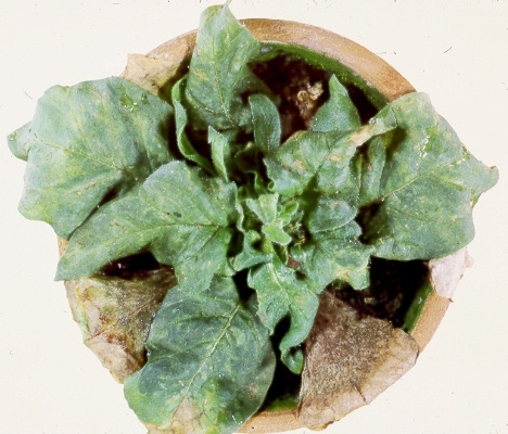
N. clevelandii plant systemically infected with an NM-type isolate, showing severe symptoms.

Necrotic local lesions in inoculated leaf of Chenopodium amaranticolor.

L particles arranged radially around a mitochondrion in a N. clevelandii leaf cell. Bar represents 200 nm.

Section of X-body (X) adjacent to the nucleus (N) in a leaf hair cell of N. clevelandii infected with an NM-type isolate. Bar represents 500 nm.
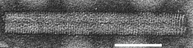
Particle negatively stained with uranyl formate + formic acid, showing transverse striations indicative of helical structure. Bar represents 50 nm.

Particles negatively stained with 2% phosphotungstate. Bar represents 100 nm.
References list for DPV: Pepper ringspot virus (347)
- Abou Haidar & Hirth, Virology 76: 173, 1977.
- Ball, Minson & Shih, Nature, Lond., New Biol. 246: 206, 1973.
- Barker & Harrison, J. gen. Virol. 35: 135, 1977.
- Bergh, Koziel, Huang, Thomas, Gilley & Siegel, Nucleic Acids Res. 13: 8507, 1985.
- Bisaro & Siegel, Virology 107: 194, 1980.
- Camargo, Kitajima & Costa, Phytopath. Z. 64: 282, 1969.
- Chagas, Flores & Caner, Biológico 35: 271, 1969.
- Cooper & Mayo, J. gen. Virol. 16: 285, 1972.
- Costa & Kitajima, Revta Soc. bras. Fitopatol. 2: 25, 1968.
- Fritsch, Mayo & Hirth, Annls Microbiol. (Inst. Pasteur) A 127: 93, 1976.
- Frost, Harrison & Woods, J. gen. Virol 1: 57, 1967.
- Harrison & Klug, Virology 30: 738, 1966.
- Harrison & Nixon, J. gen. Microbiol. 21: 569, 1959.
- Harrison & Roberts, J. gen. Virol. 3: 121, 1968.
- Harrison & Woods, Virology 28: 610, 1966.
- Harrison, Stefanac & Roberts, J. gen. Virol. 6: 127, 1970.
- Kitajima & Costa, J. gen. Virol. 4: 177, 1969.
- Kitajima, Oliveira & Costa, Bragantia 28: 1, 1969.
- Kubo, Harrison, Robinson & Mayo, J. gen. Virol. 27: 293, 1975.
- Kurppa, Jones, Harrison & Bailiss, Ann. appl. Biol. 98: 243, 1981.
- Lister, Fedn Proc. Fedn Am. Socs exp. Biol. 28: 1875, 1969.
- Lister & Bracker, Virology 37: 262, 1969.
- Mayo, Intervirology 17: 240, 1982.
- Mayo & Cooper, J. gen. Virol. 18: 281, 1973.
- Mayo, Fritsch & Hirth, Virology 69: 408, 1976.
- Minson & Darby, J. gen. Virol. 19: 253, 1973.
- Roberts & Mayo, J. Ultrastruct. Res. 71: 49, 1980.
- Robinson, J. gen. Virol. 35: 37, 1977.
- Robinson, J. gen. Virol. 64: 657, 1983.
- Robinson & Harrison, J. gen. Virol. 66: 171, 1985.
- Salomão, Proc. 2nd Cong. int. di Studi sul Carciofo, Bari. 831, 1973.
- Silberschmidt, Phytopath. Z. 46: 209, 1962-63.
- Tollin & Wilson, J. gen. Virol. 13: 433, 1971.
- Van Belkum, Cornelissen, Linthorst, Bol, Pleij & Bosch, Nucleic Acids Res. 15: 2837, 1987.