Details of DPV and References
DPV NO: 354 December 1989
Family: Alphaflexiviridae
Genus: Potexvirus
Species: Potato virus X | Acronym: PVX
This is a revised version of DPV 4
Potato virus X
Renate Koenig Biologische Bundesanstalt für Land- und Forstwirtschaft, Institut für Viruskrankheiten der Pflanzen, Messeweg 11, D-3300 Braunschweig, Germany
D.-E. Lesemann Biologische Bundesanstalt für Land- und Forstwirtschaft, Institut für Viruskrankheiten der Pflanzen, Messeweg 11, D-3300 Braunschweig, Germany
Contents
- Introduction
- Main Diseases
- Geographical Distribution
- Host Range and Symptomatology
- Strains
- Transmission by Vectors
- Transmission through Seed
- Transmission by Grafting
- Transmission by Dodder
- Serology
- Nucleic Acid Hybridization
- Relationships
- Stability in Sap
- Purification
- Properties of Particles
- Particle Structure
- Particle Composition
- Properties of Infective Nucleic Acid
- Molecular Structure
- Genome Properties
- Satellite
- Relations with Cells and Tissues
- Ecology and Control
- Notes
- Acknowledgements
- Figures
- References
Introduction
- Described by
Smith (1931).
Selected synonym:
- Potato mild mosaic virus (Rev. appl. Mycol. 22: 367)
-
A virus with filamentous particles c. 515 x 13 nm and a monopartite positive-sense single-stranded RNA genome. It infects species in the Solanaceae and in 15 other families. It is readily sap-transmissible and is transmitted in nature mainly by mechanical contact. Occurs world-wide in potato-growing areas.
Main Diseases
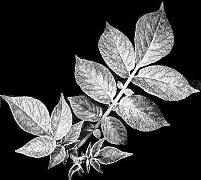
In potato the virus causes a mild mosaic (Fig. 1) or is latent; yield losses are 10 to 20% (Bode, 1968). In tobacco it causes mottling or necrotic spotting, in tomato it causes mosaic and slight stunting. In all three hosts the severity of symptoms greatly increases in mixed infections with other viruses, e.g. potato virus Y (Bode, 1968; Bode & Klinkowski, 1968; Klinkowski & Uschdraweit, 1968).
Geographical Distribution
World-wide in potato growing areas.
Host Range and Symptomatology
The virus infects more than 240 species in 16 families; the majority of hosts are in the Solanaceae (Purcifull & Edwardson, 1981).
Diagnostic species
- Nicotiana tabacum
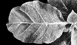
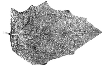
and other Nicotiana species. The first infected leaves often develop necrotic ringspots (Fig. 2); leaves infected later show chlorotic or necrotic mottling, mosaic or veinal chlorosis (Fig. 3).- Datura stramonium. Small chlorotic rings followed by mottling, veinal chlorosis or veinal necrosis (Fig. 4).
Propagation species
- Cultivars of Nicotiana tabacum and other Nicotiana spp.
Assay species
- Gomphrena globosa
is a useful local lesion host for most strains except for the resistance- breaking strain HB (Moreira et al., 1980).
Strains
Many minor variants can be distinguished, mainly by the symptoms they give in tobacco. Groups of strains have been distinguished on the basis of (i) reactivity with polyclonal antisera (Matthews, 1949) or monoclonal antibodies (Torrance et al., 1986), or (ii) different thermal inactivation points (Köhler, 1962), or (iii) infectivity for potato cultivars carrying different resistance genes (Cockerham, 1955; 1970; see Ecology and Control). There is no strong correlation between the groupings based on these different parameters. A strain (HB) which breaks the resistance of several potato clones has been described (Moreira et al., 1980).
Transmission by Vectors
Although the virus is spread mainly by mechanical contact (see Ecology and Control) transmission has been reported by the fungus Synchytrium endobioticum (Nienhaus & Stille, 1965) and the grasshoppers Melanoplus differentialis (Walters, 1952) and Tettigonia viridissima (Schmutterer, 1961). The grasshoppers probably transmit the virus on their mouthparts.
Transmission through Seed
None reported.
Transmission by Dodder
Results inconclusive; the virus infects some dodder species (Schmelzer, 1956).
Serology
The virus is strongly immunogenic and can be detected by a variety of serological tests. Time-resolved fluoroimmunoassay (Sinijärv et al., 1988) is among the most sensitive tests (detection limit 5 pg/ml). By means of monoclonal antibodies Koenig & Torrance (1986) identified three antigenic determinants on the viral coat protein: one is found on the protruding N-terminus of undenatured virus particles only; another is found, also on undenatured virus particles, when the N-terminus has been removed by proteolysis; and a third determinant is hidden and becomes exposed only after partial or complete denaturation of the virus particles. A fourth determinant described by Sõber et al. (1988) is located on the protruding N-terminus and withstands denaturation with sodium dodecyl sulphate. It involves the lysine residue at position 19 (Radavsky et al., 1989).
Relationships
Cross-protection among serotypes may be complete or partial (Matthews, 1949).
The virus is the type member of the potexvirus group and is serologically distantly related to several other potexviruses, e.g. cactus X, clover yellow mosaic, cymbidium mosaic, hydrangea ringspot, narcissus mosaic, papaya mosaic and white clover mosaic viruses (Koenig & Lesemann, 1978). In genome organization and amino acid sequences of structural and nonstructural proteins, potato virus X closely resembles other potexviruses but its different gene products also have more distant amino acid homologies with those of viruses belonging to various other taxonomic groups (see Genome Properties).
Stability in Sap
In tobacco sap, the thermal inactivation point (10 min) is between 68° and 74°C depending on the strain, the dilution end-point is between 10-5 and 10-6, and infectivity is retained at 20°C for several weeks (Bode, 1968).
Purification
Mince 100 g infected tobacco leaves in 100 ml of a solution containing 0.5 M boric acid adjusted to pH 7.8 with NaOH. Adjust the pH of the expressed sap to 6.5, add 0.2% (w/v) ascorbic acid and 0.2% (w/v) sodium sulphite and centrifuge at low speed. To 1 vol. of the supernatant fluid add 0.15 vol. 0.5% silver nitrate and leave at room temperature for 2-3 h. Centrifuge at low speed and add polyethylene glycol M. Wt 6000 to the supernatant fluid to a final concentration of 4% (w/v). Leave at 4°C overnight. Centrifuge at low speed and resuspend the pellets in a solution containing 3% urea and 0.1 % (v/v) mercaptoethanol in 0.5 M boric acid adjusted to pH 7.8 with NaOH. Subject the virus to one cycle of differential centrifugation and purify further by caesium chloride density gradient centrifugation. This method yields virus which contains the intact coat protein. Virus particles which partially or completely lack an N-terminal portion of the coat protein are obtained when the expressed sap is adjusted to pH 5.5 and the particles are left in prolonged contact with the sap in the presence of reducing agents (Koenig et al., 1978).
Properties of Particles
Sedimentation coefficient (s20,w) at infinite dilution: 117.7 S.
Particle weight: c. 35 x 106, calculated from data given by Huisman et al. (1988) and Tollin & Wilson (1988).
Isoelectric point: pH 4.4 (Kim & Hooker, 1959).
Partial specific volume: 0.73 cm3/g (Lauffer & Cartwright, 1952).
Electrophoretic mobility: -4 x 10⁻⁶ cm2 s-1 V-1 (Bawden & Kleczkowski, 1959).
A260 nm(0.1%, 1 cm): 2.97 (Paul, 1959).
A260/A280: 1.20 (Paul, 1959).
Particle Structure
The particles are flexuous filaments c. 515 nm long with a diameter of c. 13 nm (Brandes, 1964). About 1270 identical protein subunits are arranged in a helix with a pitch of c. 3.4 to 3.6 nm depending on the humidity; the true repeat of the helix is 8 turns with 8.875 subunits per turn (Tollin & Wilson, 1988). The nucleic acid seems to be located at a radius of c. 3.5 nm (Varma et al., 1968). An axial canal with a radius of c. 1.7 nm can be seen in end-on views of very short lengths of the particles but is not visible in micrographs of complete particles (Varma et al., 1968). In well-resolved micrographs the particles show a regular surface pattern of cross-banding and longitudinal files of subunits (Fig. 8).
Particle Composition
Nucleic acid: Linear single-stranded positive-sense RNA. One molecule per particle, about 6% of particle weight, calculated from data by Huisman et al. (1988) and Tollin & Wilson (1988). The nucleotide sequence (6435 bases excluding the 3' poly(A) tail) has been determined (Huisman et al., 1988; Skryabin et al., 1988).
Coat protein: Approximately 94% of particle weight; c. 1270 identical subunits of M. Wt 25,080 (Tollin & Wilson, 1988; Huisman et al., 1988). The coat protein in the intact virus can be partially degraded at the N-terminus by reducing agent-dependent proteases and trypsin, and at the C-terminus by reducing agent-independent proteases that contaminate some virus preparations (Koenig et al., 1978).
Genome Properties
The genomic RNA of the virus
(Fig. 9)
has a 5' m7GpppG cap
(Sonnenberg et al., 1978),
a 3' poly(A) tail
(Morozov et al., 1987)
and five open reading frames (ORFs)
(Huisman et al., 1988;
Skryabin et al., 1988).
ORF 1 (bases 85-4453) is preceded by
a 84-base 5' leader sequence; ORF 3 (5147-5492) partially overlaps with
ORF 2 (4486-5164) and ORF
4 (5427-5637); ORF 5 (5650-6361) is followed by an untranslated region of 76 bases.
ORF 1-5 code,
respectively, for proteins of M. Wt 166 K, 25 K, 12 K, 8 K and 25 K (coat protein).
Homologies with
gene products of other viruses (see below) suggest that ORF 1 encodes an RNA replicase,
the central
regions of ORF 1 and ORF 2 encode NTP-binding helicase(s) and
ORF 3 and 4 encode membrane-bound
proteins which may have a transport function
(Skryabin et al., 1988).
Infected plants contain 2 major (0.9 and 2.1 kb) and 4 minor (1.4, 1.8, 3.0 and 3.6 kb)
subgenomic RNAs,
and double-stranded analogues to most of them. The 0.9 kb subgenomic RNA is the
coat protein messenger
(Dolja et al., 1987).
In vitro translation of the genomic RNA yields two major
polypeptides of 145 and 180 K; they share common amino acid sequences
but the 180 K protein is not
a precursor of the 145 K protein
(Wodnar-Filipowicz et al., 1980).
The genome organization of potato virus X (Fig. 9) and the relative positions of the individual cistrons (Huisman et al., 1988; Skryabin et al., 1988) are identical to those of white clover mosaic (Forster et al., 1988) and narcissus mosaic (Zuidema et al., 1989) potexviruses and similar to those of carlaviruses (MacKenzie et al., 1989). The triple gene block formed by ORF 2, 3 and 4 (Fig. 9) resembles a probably functionally similar triple gene block on the RNA-2 molecules of beet necrotic yellow vein furovirus and barley stripe mosaic hordeivirus (Morozov et al., 1989). The amino acid sequences of all five ORFs of potato virus X show pronounced homologies with those of the corresponding genome regions of other potexviruses (Forster et al., 1988; Zuidema et al., 1989). The amino acid sequence encoded by ORF 1 has the -GxxGxGKT/S- and -GDD- motifs which are found in the putative replicases of many other plant and animal viruses, and the amino acid sequences surrounding these two blocks show strong homologies with those found for tymoviruses (Morozov et al., 1989; Ding et al., 1990). On the other hand amino acid sequences encoded by ORF 2 and 3 have strong homologies with putative gene products of carlaviruses, beet necrotic yellow vein furovirus and barley stripe mosaic hordeivirus (Huisman et al., 1988; MacKenzie et al., 1989; Rupasov et al., 1989). The coat protein, gene product of ORF 5, has homologies with the coat proteins of carlaviruses (MacKenzie et al., 1989; Rupasov et al., 1989), but surprisingly also with those of potyviruses (Morozov et al., 1987) and to a lesser extent also with that of barley yellow mosaic virus (Kashiwazaki et al., 1989). Potyviruses are considered to be ‘picorna-like’, whereas the other viruses to which the potato virus X genome has homologies are ‘sindbis-like’ (Goldbach & Wellink, 1988). As with other viruses, different parts of the genome probably have different phylogenetic origins (Morozov et al., 1989; Ding et al., 1990).
Relations with Cells and Tissues
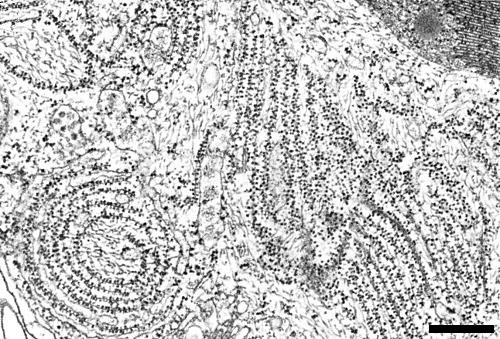
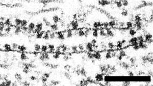
The virus particles occur, mostly in large aggregates, in the cytoplasm. The aggregates usually show a fibrous structure (Fig. 5) resulting from the parallel arrangement of the particles (for review see Lesemann, 1988a), but may also appear banded when the particle ends are ordered in register (Appiano & Pennazio, 1972; Christie & Edwardson, 1977). The aggregates may fill the greater part of the cells. The virus is the only potexvirus known to induce in the cytoplasm the formation of cytoplasmic laminated inclusion components (LIC). The LIC are proteinaceous sheets 3 nm thick which may or may not be covered on both sides with dark staining beads 11 to 14 nm in diameter (Fig. 6, Fig. 7) (Kozar & Sheludko, 1969; Shalla & Shepard, 1972). The LIC are not related serologically to viral coat protein and the beads are different from ribosomes (Shalla & Shepard, 1972). The function of the LIC is unknown. In protoplasts they appear later than the first virus particles (Honda et al., 1975). The virus also induces a proliferation of the endoplasmic reticulum and the formation of tonoplast-associated vesicles c. 100 nm in diameter in the vacuoles. The vesicles contain fibrillar structures resembling dsRNA structures as seen with many sindbis-like ss(+)RNA viruses (for review see Lesemann, 1988b).
Ecology and Control
The virus is highly infectious and is spread by mechanical contact, especially on agricultural and horticultural equipment. In potato crops, the most effective means of protection is the use of resistance conferred by major genes. Several strain-specific genes (e.g. Nx, Nb) cause hypersensitivity, and virus isolates fall into four groups according to their response to these genes (Cockerham, 1955). Two further genes (RXadg and RXacl) confer a more comprehensive extreme resistance to all four groups of strains (Cockerham, 1970). Cultivars possessing one or more of these various forms of resistance have been widely grown for many years and are effective in controlling the virus. Although a strain (HB) able to break all known forms of resistance was identified from South America (Moreira et al., 1980), resistance-breaking strains do not seem to have become prevalent elsewhere. Attention is now turning to the introduction of novel forms of resistance through the use of genetic engineering techniques. In transgenic potato plants expressing the viral coat protein gene, the development of symptoms was delayed and the accumulation of virus was greatly reduced (Hoekema et al., 1989). In transgenic tobacco plants expressing the viral coat protein gene, both the number of local lesions in the inoculated leaves and the virus concentration in the systemically infected leaves were reduced. The protection was effective not only against the virus, but also against the viral RNA suggesting that it is not, or not entirely, based on a prevention of virus uncoating (Hemenway et al., 1988).
Acknowledgements
The senior author’s work on potato virus X was supported by the Deutsche Forschungsgemeinschaft.
Figures
References list for DPV: Potato virus X (354)
- Appiano & Pennazio, J. gen. Virol. 14: 237, 1972.
- Bawden & Kleczkowski, Virology 7: 375, 1959.
- Bode, in Pflanzliche Virologie, Vol. II, part 1: p. 31, ed. M. Klinkowski, 457 pp., Berlin: Akademie-Verlag, 1968.
- Bode & Klinkowski, in Pflanzliche Virologie, Vol. II, part 1: p. 161, ed. M. Klinkowski, 457 pp., Berlin: Akademie-Verlag, 1968.
- Brandes, Mitt. biol. BundAnst. Ld.- u. Forstw. 110: 130 pp., 1964.
- Christie & Edwardson, Monogr. Ser. Fla agric. Exp. Stn 9: 155 pp., 1977.
- Cockerham, Proc. 2nd Conf. Potato Virus Diseases, Lisse-Wageningen, 1954: 89, 1955.
- Cockerham, Heredity 25: 309, 1970.
- Ding, Keese & Gibbs, J. gen. Virol. 71: 925, 1990.
- Dolja, Grama, Morozov & Atabekov, FEBS Lett. 214: 308, 1987.
- Forster, Bevan, Harbison & Garner, Nucleic Acids Res. 16: 291, 1988.
- Goldbach & Wellink, Intervirology 29: 254, 1988.
- Hemenway, Fang, Kaniewski, Chua & Turner, EMBO J. 7: 1273, 1988.
- Hoekema, Huisman, Molendijk, van den Elzen & Cornelissen, Biotechnology 7: 273, 1989.
- Honda, Kajita, Matsui, Otsuki & Takebe, Phytopath. Z. 84: 66, 1975.
- Huisman, Linthorst, Bol & Cornelissen, J. gen. Virol. 69: 1789, 1988.
- Kashiwazaki, Hayano, Minobe, Omura, Hibino & Tsuchizaki, J. gen. Virol. 70: 3015, 1989.
- Kim & Hooker, Amer. Potato J. 36: 297, 1959.
- Klinkowski & Uschdraweit, in Pflanzliche Virologie, Vol. II, part 2: p. 1, ed. M. Klinkowski, 460 pp., Berlin: Akademie-Verlag, 1968.
- Koenig & Lesemann, CMI/AAB Descr. Pl. Viruses 200, 5 pp., 1978.
- Koenig & Torrance, J. gen. Virol. 67: 2145, 1986.
- Koenig, Tremaine & Shepard, J. gen. Virol. 38: 329, 1978.
- Köhler, Phytopath. Z. 44: 189, 1962.
- Kozar & Sheludko, Virology 38: 220, 1969.
- Lauffer & Cartwright, Arch. Biochem. Biophys. 38: 371, 1952.
- Lesemann, in The Plant Viruses Vol. 4, p. 179, ed. R. G. Milne, New York: Plenum Press, 1988a.
- Lesemann, Acta Hort. 234: 289, 1988b.
- MacKenzie, Tremaine & Stace-Smith, J. gen. Virol. 70: 1053, 1989.
- Matthews, Ann. appl. Biol. 36: 480, 1949.
- Moreira, Jones & Fribourg, Ann. appl. Biol. 95: 93, 1980.
- Morozov, Lukashewa, Chernow, Skryabin & Atabekov, FEBS Lett. 213: 438, 1987.
- Morozov, Dolja & Atabekov, J. molec. Evol. 29: 52, 1989.
- Nienhaus & Stille, Phytopath. Z. 54: 335, 1965.
- Paul, Arch. Mikrobiol. 32: 416, 1959.
- Purcifull & Edwardson, in Handbook of Plant Virus Infections and Comparative Diagnosis, p. 628, ed. E. Kurstak, Amsterdam: Elsevier/North-Holland, 1981.
- Radavsky, Viter, Turova, Zeitseva, Järvrekülg, Saarma, Grebenshchikov & Baratova, Bioorganicheskaya Chimia 15: 619, 1989.
- Rupasov, Morozov, Kanyuka & Zavriev, J. gen. Virol. 70: 1861, 1989.
- Schmelzer, Phytopath. Z. 28: 1, 1956.
- Schmutterer, Z. angew. Ent. 47: 277, 1961.
- Shalla & Shepard, Virology 49: 645, 1972.
- Sinijärv, Järvekülg, Andreeva & Saarma, J. gen. Virol. 69: 991, 1988.
- Skryabin, Kraev, Morozov, Rozanov, Chernov, Lukashewa & Atabekov, Nucleic Acids Res. 16: 10929, 1988.
- Smith, Proc. R. Soc. Ser. B 109: 251, 1931.
- Sõber, Järvekülg, Toots, Radavsky, Villems & Saarma, J. gen. Virol. 69: 1799, 1988.
- Sonnenberg, Shatkin, Riccardi, Rubin & Goodman, Nucleic Acids Res. 5: 2501, 1978.
- Tollin & Wilson, in The Plant Viruses Vol. 4, p. 51, ed. R. G. Milne, New York: Plenum Press, 1988.
- Torrance, Larkins & Butcher, J. gen. Virol. 67: 57, 1986.
- Varma, Gibbs, Woods & Finch, J. gen. Virol. 2: 107, 1968.
- Walters, Phytopathology 42: 355, 1952.
- Wodnar-Filipowicz, Skrzeczkowski & Filipowicz, FEBS Lett. 109: 152, 1980.
- Zuidema, Linthorst, Huisman, Asjes & Bol, J. gen. Virol. 70: 267, 1989.
