Details of DPV and References
DPV NO: 390 April 2002
Family: Caulimoviridae
Genus: Badnavirus
Species: Banana streak virus | Acronym: BSV
Banana streak virus
Andrew D.W. Geering Agency for Food and Fibre Sciences, Queensland Horticulture Institute, Department of Primary Industries, 80 Meiers Road, Indooroopilly 4068, Queensland, Australia
John E. Thomas Agency for Food and Fibre Sciences, Queensland Horticulture Institute, Department of Primary Industries, 80 Meiers Road, Indooroopilly 4068, Queensland, Australia
Contents
- Introduction
- Main Diseases
- Geographical Distribution
- Host Range and Symptomatology
- Strains
- Transmission by Vectors
- Transmission through Seed
- Transmission by Grafting
- Transmission by Dodder
- Serology
- Nucleic Acid Hybridization
- Relationships
- Stability in Sap
- Purification
- Properties of Particles
- Particle Structure
- Particle Composition
- Properties of Infective Nucleic Acid
- Molecular Structure
- Genome Properties
- Satellite
- Relations with Cells and Tissues
- Ecology and Control
- Notes
- Acknowledgements
- Figures
- References
Introduction
Banana streak disease was first described as 'la mosaïque à tirets' by Yot-Dauthy & Bové (1966).
Banana streak virus (BSV) was first described by Lockhart (1986).
BSV has bacilliform-shaped particles, ca 30 x 130-150 nm in size (Fig. 1), and a circular dsDNA genome of ca 7.4 kb (Harper & Hull, 1998; Lockhart & Jones, 1999). The virus is transmitted by mealybugs, but not by mechanical inoculation of sap extracts. The only known hosts of BSV are within the family Musaceae, and the virus probably occurs wherever bananas are cultivated. Symptom expression is sporadic but fruit yields are often significantly reduced (Lassoudièrre, 1974; Dahal et al., 2000; Daniells et al., 2001). BSV is a limiting factor in the development of new Musa hybrids by breeding programs across the world. Infection in these new hybrids is often thought to originate from viral sequences that are integrated in the host's nuclear genome (Ndowora et al., 1999; Harper et al., 1999b).
Main Diseases
BSV causes banana streak disease (Lockhart, 1986). Disease severity is very variable, and probably depends on environmental conditions, as well as on host and virus genotypes. Mixed infections of BSV and Banana mild mosaic virus are also common (C. F. Gambley, L. A. McMichael & J. E. Thomas, unpublished data), and the nature of interactions between these two viruses is unknown. The most characteristic foliar symptoms of infection are chlorotic streaks (Fig. 2 and Fig. 3), which become necrotic with time (Fig. 4). The leaf lamina may also be narrower, thicker and become torn. Other symptoms include stunting of the plant, constriction of the bunch on emergence (choking), altered phyllotaxis (leaves arranged in a single vertical plane instead of the normal spiral pattern), and detachment and splitting of the outer leaf sheaths of the pseudostem (Fig. 5). In severe instances, cigar leaf necrosis (Fig. 6), internal necrosis (Fig. 7) and collapse of the pseusodstem is observed (Lockhart, 1995). Infection can cause a delay in bunch emergence and harvest, a reduction in bunch weight, and shorter and abnormally shaped fruit with a thinner fruit peel that is prone to splitting (Lockhart & Jones, 1999; Daniells et al., 2001). In some instances, bunches burst out midway up the pseudostem.
Foliar symptoms are periodic in expression; in one study, only 45-51% of the leaves produced in a single plant generation showed symptoms (Daniells et al., 2001). Periods of symptom expression do follow distinct patterns, but the underlying reasons for these patterns are unclear. In Nigeria, greater symptom expression was observed when plants were grown at 22°C in a growth cabinet than in a screenhouse at 28-35°C (Dahal et al., 1998a) and in the cooler wet season compared with the hot dry season (Dahal et al., 1998b, 2000). However, contrasting correlations with temperature were observed in Morocco and Australia, where greater symptom expression occurred during hotter times of the year (Lockhart, 1986; Daniells et al., 2001). Daniells et al. (2001) noted that over two plant generations, symptom expression peaked at the time of bunch initiation, suggesting that plant developmental stage may be a factor in symptom expression.
Lockhart & Jones (1999) proposed that most fruit abnormalities occur when flushes of symptoms coincide with the time of bunch initiation, but this hypothesis is not supported by studies in Australia (Daniells et al., 2001). Daniells et al. (2001) did observe greater effects of infection on fruit yield in the ratoon crop compared with the newly planted crop, suggesting that as plantings become older, and/or overall environmental stresses greater, infection is more detrimental to yield.
BSV-GF has been detected in Ensete ventricosum (a close relative of bananas) from Kenya with streak symptoms (A. D. W. Geering, J. N. Parry, I. Buddenhagen & J. E. Thomas, unpublished data). Some badnavirus isolates from diseased sugarcane and banana plants are closely related serologically (Lockhart & Autrey, 1988) and it is possible that there is movement of virus between these two crops. Sugarcane bacilliform virus has been experimentally transmitted to bananas by agro-inoculation and by mealybugs and, in this host, elicits symptoms typical of BSV infection (Bouhida et al., 1993; Lockhart, 1995).
Geographical Distribution
The virus probably occurs wherever bananas are grown and has been found in Africa, Europe (Canary Islands and Madeira), Asia, North (Florida), Central and South America, Australasia, and the Pacific Islands (Diekmann & Putter, 1996; Lockhart & Jones, 1999).
Host Range and Symptomatology
Experimentally, BSV has been transmitted only to plants within the family Musaceae using mealybug vectors, but never by mechanical inoculation of sap extracts (Lockhart & Jones, 1999). Many different banana genotypes are susceptible to BSV infection, including Musa acuminata (Musa AA and AAA groups) and M. balbisiana (Musa BB group) types, as well as hybrids of these two species (Musa AAB, ABB, ABBB groups) (Harper et al., 1999a; Lockhart & Jones, 1999; Geering et al., 2000). Ensete ventricosum is both an experimental and natural host of this virus (Lockhart, 1995; A. D. W. Geering, J. N. Parry, I. V. Buddenhagen & J. E. Thomas, unpublished data).
Strains
The taxonomy of BSV is currently under revision. Three virus isolates from Australia, namely BSV-Mys (from cv. Mysore), BSV-GF (from cv. Goldfinger) and BSV-IM (from CIRAD hybrids IRFA 909, 910, 914) are sufficiently different from each other and from BSV-OL (from cv. Obino l'Ewai, Nigeria) to be considered distinct virus species (Geering et al. 2000; A. D. W. Geering, J. N. Parry & J. E. Thomas, unpublished data). An additional virus isolate from Australia, namely BSV-Cav (from cv. Williams), is possibly a strain of BSV-OL (Geering et al., 2000). Lockhart & Olszewski (1993) examined five different virus isolates, namely BSV-Mad/DC (from cv. Dwarf Cavendish, Madeira), BSV-MA (from cv. Grand Nain, Morocco), BSV-Mys/T (from cv. Mysore, Trinidad), BSV-DC/Mdg from cv. Dwarf Cavendish, Madagascar) and BSV-RW (from Rwanda). Preliminary studies suggest that BSV-RW is sufficiently different from the previously mentioned virus isolates to be regarded as a separate species, but BSV-Mys and BSV-Mys/T are synonymous (A. D. W. Geering & B. E. L. Lockhart, unpublished data). The relationships of BSV-Mad/DC, BSV-DC/Mdg and BSV-MA to the other virus isolates have yet to be examined.
Transmission by Vectors
BSV is transmitted by mealybugs, most probably in a semi-persistent manner (Lockhart & Jones, 1999; Kubiriba et al., 2001b). In glasshouse experiments, Planococcus citri and Saccharicoccus sacchari, both known to colonise banana, have been shown to be capable of transmitting BSV (Lockhart, 1995). In surveys of Uganda banana farms, P. citri was absent and Dysmicoccus brevipes and S. sacchari were the most abundant mealybug species present (Kubiriba et al., 2001b). In this study, a significant positive correlation was found between the incidence of BSV and the abundance of D. brevipes. However, the ability of D. brevipes to transmit Ugandan isolates of BSV awaits confirmation. Kubiriba et al. (2001b) transmitted a badnavirus to bananas using mealybugs collected directly from field-grown pineapples. However, the identity of the mealybugs was not confirmed, and it is possible that Pineapple bacilliform virus instead of BSV was transmitted to the bananas.
Transmission through Seed
A report of seed transmission of BSV (Daniells et al., 1995) must now be considered inconclusive, as the possibility that infection in the seedlings arose from expression of integrated viral DNA cannot be excluded.
Serology
BSV is moderately immunogenic. An antiserum prepared in a rabbit had a titre of 1/512 in a gel double diffusion test using partially purified antigens (Lockhart, 1986). Other antisera have been prepared, including polyclonal antisera in rabbits (Lockhart & Olszewski, 1993; Ndowora, 1998), mice (Thottappilly et al., 1998) and chickens (Hughes, 1998; B. E. L. Lockhart, personal communication), and monoclonal antibodies (Ndowora, 1998). These antisera are suitable for use in a variety of ELISA formats and ISEM (Ndowora, 1998; Thottappilly et al., 1998).
Isolates of BSV are serologically diverse, and individual antisera often fail to detect some BSV isolates (Lockhart & Olszewski, 1993; Ndowora, 1998). A useful antiserum for broad spectrum detection of BSV isolates was prepared using BSV-Mys and a mixture of 32 isolates of Sugarcane bacilliform virus (Ndowora, 1998).
Relationships
Phylogenetic analyses using the conserved reverse transcriptase/RNase H region of the ORF III polyprotein suggests that banana streak disease is caused by several distinct badnaviruses (Geering et al., 2000; G. Harper, personal communication). The genetic distances between BSV-OL, -GF, -Mys and -IM are sufficiently great for these viruses to be recognised as distinct species, although presently they are still referred to as BSV isolates. These four isolates do cluster together in a phylogenetic tree, but there are other isolates, such as BSV-RW, that are much more closely related to Sugarcane bacilliform virus (SCBV) than to any of the previously mentioned isolates. SCBV also infects a range of monocot hosts, including banana in which it elicits typical banana streak disease symptoms (Bouhida et al., 1993; Lockhart, 1995). Badnavirus isolates from sugarcane are genetically diverse (Braithwaite et al., 1997), and it is probable that there is some interchange of viruses between this crop and bananas.
Stability in Sap
There are no reports of infective virus being retrieved from preserved virus cultures. However, freeze-dried or frozen (-70°C) leaf tissue and sap extracts from infected plants stored under liquid nitrogen, and partially purified virus minipreps frozen at -20°C, have all served as sources of virus for ELISA, PCR and ISEM tests (A. D. W. Geering, J. N. Parry, L. A. McMichael & J. E. Thomas, unpublished data).
Purification
Banana leaf tissue with strong disease symptoms generally contains the highest virus titres (Lockhart, 1986; Dahal et al., 1998b; Daniells et al., 2001). Several methods have been reported for the purification of BSV.
Lockhart (1986). Extract tissue in 2 vol. 0.05 M Tris-citrate buffer, pH 7.4, containing 0.5% (w/v) Na2SO3, 1% (w/v) PVP (mol. wt 40,000) and 1% Triton X-100. Squeeze extract through muslin, emulsify for 20 sec with 25% (v/v) chloroform, and then centrifuge at 10,000 g for 10 min. The aqueous phase is ultracentrifuged at 136,000 g for 1 h, and the pellets resuspended in 0.01 M phosphate buffer, pH 7.2. Further purification is achieved by one or two cycles of centrifugation for 4.5 h at 116,000 g in a preformed 0-30% CsCl gradient containing 10% (w/w) sucrose.
Thottappilly et al. (1998), modified from Lockhart (1986). Freeze leaf tissue with liquid nitrogen and pulverise. Thaw the powder in 2-3 vol. 0.2 M sodium phosphate buffer pH 6.0, containing 10 mM EDTA, 0.5% Na2SO3, 0.5% 2-mercaptoethanol, 2% PVP, 4% PEG; 1 ml Antifoam A (Sigma) is added per 2 L extraction buffer. Squeeze extract through cheesecloth, and centrifuge at 12000 g for 20 min. To the supernatant fluid, add PEG to a final concentration of 6% (w/v), and stir for 1-2 h at 4°C. The PEG precipitate is collected by centrifuging at 12,000 g for 20 min and resuspended by stirring for 10-15 min in 10 mM sodium phosphate buffer, pH 7.2, containing 1.5% NaCl, 1 mM MgCl2, and 2% Triton X-100. The extract is then clarified by low speed centrifugation, and the virus pelleted by ultracentrifugation at 105,000 g for 90 min. The resuspended pellets are emulsified with an equal volume of chloroform, the aqueous phase recovered, and the virus further purified by centrifuging for 6 h in a Cs2SO4 step gradient (15, 22.5 and 30% w/v) in 10 mM Tris- HCl buffer pH 7.4.
Geering et al. (2000), based on Adomako et al. (1983). Leaf tissue is extracted in 8 vol. 0.05 M sodium phosphate buffer pH 6.1, containing 5 mM DIECA, 0.2% (v/v) thioglycerol, 0.5% (w/v) PEG 6,000, and 0.5% (v/v) Celluclast (Novo Nordisk, Denmark). The extract is stirred at room temperature for 5 h, then left overnight at 5°C. After squeezing through cheesecloth and centrifuging at 3,000 g for 20 min, PEG (mol. wt 6,000) and NaCl (9.5% w/v and 0.2 M final concentration, respectively) is added. The extract is stirred for 15 min, then left at room temperature for 3 h, and the PEG precipitate collected by centrifuging at 10,000 g for 20 min. The pellet is resuspended in 1/30 original extraction volume of 0.05 M sodium phosphate buffer pH 6.8, 0.2 M NaCl, 0.1% Na2SO3, 5 mM EDTA (buffer B) and clarified by centrifuging at 8,000 g for 20 min. The pellet is re-extracted as above, the two supernatant fluids combined and Celite (2 g/30 g starting tissue) added, mixed and collected on filter paper in a Buchner funnel under gentle suction. NaCl and PEG, at final concentrations of 0.2 M and 7%, respectively, are added to the filtrate, and the mixture placed on a 4 cm bed of Celite (equilibrated with buffer B) in a 2 cm diameter column. The column is eluted with stepwise additions of 25 ml aliquots of buffer B containing 5%, 3%, 1%, then 0% of PEG (mol. wt 6,000). The eluates from each step are clarified by centrifuging at 7,000 g for 10 min at 10°C, and the supernatant fluids centrifuged at 50,000 rpm for 50 min at 5°C in a Beckman Type 70 Ti rotor. Pellets are resuspended in 0.02 M sodium citrate buffer pH 7.0 (CB) and the virus- containing fractions (determined by electron microscopy) pooled and layered onto a 10-40% sucrose density gradient in CB, and centrifuged at 35,000 rpm for 1 h at 5°C in a Beckman SW 41 rotor. Virus-containing zones are collected, concentrated by ultracentrifugation and resuspended in CB.
Properties of Particles
Purified virions have an A260/280nm ratio of 1.26 (uncorrected for light scattering) (Lockhart,1986).
Particle Structure
Particles are bacilliform in shape with a diameter of 30 nm and a length of 130-150 nm, although substantially longer particles are often observed (Fig. 1; Lockhart & Jones, 1999). The particles have an electron-dense core and the tubular portion of the particle has a structure based on an icosahedron cut across its threefold axis, with a structural repeat of 10 nm and nine rings of hexamer subunits per 130 nm length (Hull et al., 2000).
Particle Composition
Nucleic acid: Particles contain a single, circular, dsDNA molecule. The genome of BSV-OL has been completely sequenced, and is 7389 bp long (GenBank accession number AJ002234; Harper & Hull, 1998). Incomplete sequences, comprising parts of ORF III and the intergenic region, are available for BSV-Cav, BSV-Mys and BSV-GF (GenBank accession numbers AF215815, AF214005 and AF215814, respectively; Geering et al., 2000). Encapsidated badnavirus DNA typically has two discontinuities, one in each strand. With all badnaviruses, the discontinuity in the (-) sense DNA is adjacent to the 3'-end of the cytosolic initiator methionine (tRNAmeti) binding site (Medberry et al., 1990). The position of the discontinuity in the (+) sense DNA of BSV-OL has not been mapped. As is the convention, numbering of the nucleotide sequence of BSV begins at the 5'-end of the tRNAmeti binding site.
Protein: No published information available for any isolate.
Genome Properties
The genome of BSV-OL contains three ORFs, encoding two small proteins of 20.8 kDa (ORF I) and 14.5 kDa (ORF II) and a 208 kDa polyprotein (ORF III) (Harper & Hull, 1998; Fig. 8). The functions of the 20.8 and 14.5 kDa proteins have not been clearly elucidated. Studies with other badnaviruses suggest that the 14.5 kDa protein is incorporated into the virus coat protein and is possibly involved in virus assembly (Cheng et al., 1996; Stavolone et al., 2001). This protein has a short, hydrophobic and basic domain at the C-terminus of the protein, providing sequence non-specific ssRNA and dsDNA binding abilities (Jacquot et al., 1996, 1997). Additionally, the protein has a coiled-coil motif, allowing the protein to interact with itself to form tetramers, which subsequently may interact with other viral or cellular proteins (Stavolone et al., 2001). The putative components of the 208 kDa polyprotein in order from the N- to C-termini are movement protein, coat protein, aspartyl proteinase and replicase (reverse transcriptase and RNase H) (Medberry et al., 1990; Tzafrir et al., 1997; Harper & Hull, 1998). The 208 kDa polyprotein is thought to be post-translationally cleaved into functional units by the aspartyl proteinase. The sites of cleavage have not been determined.
As with Cauliflower mosaic virus, replication is assumed to begin when viral DNA is transported to the nucleus, the discontinuities in the DNA repaired, and mini-chromosomes formed by association of the covalently closed, supercoiled DNA with histone proteins (Hull, 2001). Mini-chromosomal DNA is then transcribed utilising host-encoded DNA-dependent RNA polymerase II (Hull, 2001). The putative TATA-box of the BSV-OL promoter is located at nts 7231-7237 of the BSV-OL genome, and transcription begins at either nt 7260 or 7261 (Harper & Hull, 1998). The promoters of BSV-Mys and BSV-Cav have strong, constitutive activity, especially in vascular tissue (Schenk et al., 2001). A terminally redundant RNA greater than one genome in length, termed the pregenomic RNA, is transcribed, and this RNA serves as a mRNA for translation of virus-encoded proteins. The 5' leader sequence of the pregenomic RNA of BSV-OL is 645 nucleotides long and contains several short, non-functional, ORFs (sORFs). Ribosome scanning is believed to begin at the first sORF (only six codons long), but following this, the RNA folds into a large stem-loop structure, across which the ribosome is shunted to near the beginning of ORF I (Pooggin et al., 1999). Translation of ORFs II and III is believed to occur via a "leaky" scanning process, in which preceding start codons are bypassed (Fütterer et al., 1997). In the final step of replication, the pregenomic RNA is converted back to dsDNA by action of the reverse transcriptase. Synthesis of single-stranded, (-) sense DNA, is primed by tRNAmeti. Synthesis of (+) sense DNA is thought to be primed by purine rich cleavage products left after RNase H digestion of the pregenomic RNA following synthesis of the (-) sense DNA (Medberry et al., 1990).
Relations with Cells and Tissues
BSV has been detected in the leaf lamina and midrib, pseudostem and roots of the banana plant and peel of the fruit (Dahal et al., 1998b; A. D. W. Geering & J. E. Thomas, unpublished data). There are no published reports of the intracellular location of BSV, but badnaviruses in general are restricted to the cytoplasm and are not associated with any inclusion bodies or membrane-bound structures (Hull et al., 2000). Virus titres vary greatly within and between plants growing at the same location (Dahal et al., 1998a, 1998b; Daniells et al., 2001).
An unusual attribute of BSV-OL is that its DNA is integrated in the B-genome of cultivated Musa (Ndowora et al., 1999; Harper et al., 1999b; Geering et al., 2001). There is evidence to suggest that with some Musa hybrids, infection develops de novo following activation of this integrated DNA. Integration is not an essential step in the replication cycle of the virus, and DNA has possibly become integrated over time by a process of illegitimate recombination (Ndowora et al., 1999). An integrant characterised from cv. Obino l'Ewai (Musa AAB group) contains two segments of uninterrupted viral DNA, corresponding to nts 7264 - 5810 and nts 5530 - 7363 of the BSV-OL genome, respectively, and together these comprise the complete viral genome. However, these two segments are interspersed by a 6 kb `scrambled' region containing several segments of non-contiguous and partially inverted viral DNA. In the activation model proposed by Ndowora et al. (1999), a homologous recombination occurs between the 280 bp direct duplication of DNA flanking the `scrambled' region, leading to excision of this non-functional DNA. The remaining viral DNA is then circularised and excised from the chromosome following a second homologous recombination between the 98 bp direct repeats of DNA at either end of the integrant.
Ecology and Control
It is generally regarded that the rate of spread of BSV by mealybugs is slow (Lockhart & Jones, 1999; Daniells et al., 2001; Kubiriba et al., 2001a), and that the greatest risk of dissemination of the virus is by the inadvertent use of infected planting material. Roguing is probably an adequate control measure for isolated outbreaks. However, even in widespread outbreaks, economic returns may sometimes still be acceptable, especially under intensive management conditions (Daniells et al., 2001).
In many instances, infection is thought to develop de novo from viral DNA integrated in the host's nuclear genome (Ndowora et al., 1999; Harper et al., 1999b). Integrated BSV-OL DNA is linked to the B-genome of cultivated Musa (Geering et al., 2001), and is therefore absent from the most commercially important dessert bananas in the Cavendish subgroup (Musa AAA group). The use of tissue culture to propagate bananas is known to be a trigger for expression of these integrated viral sequences (Dallot et al., 2001), but other undefined environmental conditions may also be involved. All Musa AxB hybrids are considered to have integrated BSV-OL DNA, although cultivars vary greatly in their propensity to become infected, suggesting the involvement of additional host factors in the development of infection. Cultivars Lady Finger (Musa AAB group) and Goldfinger (Musa AAAB group) have been propagated in large quantities in Australia by tissue culture, but infection in these cultivars is very rare (A. D. W. Geering & J. E. Thomas, unpublished data).
Selection of clean planting material is made difficult because infected plants can be symptomless for long periods, and virus-indexing procedures are often inadequate because of the great genetic and serological diversity of the virus, and frequently low virus titres in plants. When selecting plants for propagation, all leaves should be inspected for symptoms on several dates, including a date around the time of bunch initiation when symptom expression appears to be the greatest (Daniells et al., 2001). Plants initiated into tissue culture should be indexed for virus, and a sample of cloned plantlets also tested for virus on at least two occasions over a six month period (Diekmann & Putter, 1996).
Specific PCR assays are now available for BSV-OL, BSV-Mys, BSV-Cav and BSV-GF (Harper et al., 1999a; Geering et al., 2000), but there are additional uncharacterised isolates not detected by these assays (A. D. W. Geering & J. E. Thomas, unpublished data). Immunocapture PCR should be used to distinguish between integrated and episomal forms of the viral DNA, especially in Musa AxB hybrids (Harper et al., 1999a). ISEM is generally a more sensitive assay than ELISA (Thottappilly et al., 1998; Lockhart, 1995). For quarantine purposes, a combination of partial virus purification and immunosorbent electron microscopy using a broad spectrum BSV antiserum is currently the most reliable indexing method to detect all isolates of BSV (Diekmann & Putter, 1996). PCR assays using currently available badnavirus degenerate primers (Lockhart & Olszewski, 1993; Ahlawat et al., 1996) lack the sensitivity required for routine use, although Badna 1A and Badna 4 primers (N. E. Olszewski, personal communication) hold much more promise.
Notes
The taxonomy of the genus Badnavirus is in need of revision. For several tropical crops such as banana, sugarcane, yam and pineapple, there is a great diversity of badnavirus isolates infecting each crop (Braithwaite et al., 1997; Geering et al., 2000; Gambley et al., 2001; G. Harper & L. Kenyon, personal communication). In many cases, the relationships between badnavirus isolates from the different crops are closer than those from the same crop, and the natural host range of some of the virus isolates may include more than one crop species. Some badnavirus isolates from banana (presently called BSV) and from sugarcane (presently called SCBV) are probably identical. Currently, the only definitive way of distinguishing virus isolates is by DNA sequence analysis.
Figures

Electron micrograph of particles negatively stained with 2% uranyl acetate, pH 3.8. The bar represents 100 nm. Photograph courtesy of G. G. F. Kasdorf.

Chlorotic streaking in leaves of Musa AAB group cv. Mysore. Photograph courtesy of J. W. Daniells.
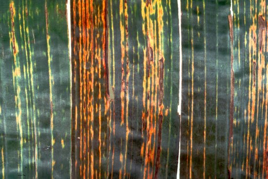
Chlorotic streaking in segments of leaf from Musa AAA group cv. Red Dacca.

Necrotic streaking in a leaf of Musa AAA group cv. Williams. Photograph courtesy of J. W. Daniells.

Leaf sheaths detached from the pseudostem of Musa AAA group cv. Williams. Photograph courtesy of J. W. Daniells.

Cigar leaf necrosis of Musa AAA group cv. Williams. Photograph courtesy of J. W. Daniells.

Internal pseudostem necrosis of Musa AAA group cv. Williams. Photograph courtesy of J. W. Daniells.

Organisation of the BSV-OL genome. The complete circle represents the double-stranded DNA genome. The inner dotted circle shows the mapping of the RNA transcript with the position of the 3' end being estimated. The outer arcs indicate the positions of the three ORFs. The figure is from Harper & Hull (1998) and reproduced courtesy of Kluwer Academic Publishers.
References list for DPV: Banana streak virus (390)
- Adomako, Lesemann & Paul, Annals of Applied Biology 103: 109, 1983.
- Ahlawat, Pant, Lockhart, Srivastava, Chakraborty & Varma, Plant Disease 80: 590, 1996.
- Bouhida, Lockhart & Olszewski, Journal of General Virology 74: 15, 1993.
- Braithwaite, Geijskes, Geering, McMichael, Thomas & Smith, Abstracts of the Pathology and Molecular Biology Workshop, International Society of Sugar Cane technologists, Umhlanga Rocks, KwaZulu-Natal, South Africa, May 1997.
- Cheng, Lockhart, Olszewski, Virology 223: 263, 1996.
- Dahal, Hughes, Thottappilly & Lockhart, Plant Disease 82: 16, 1998a.
- Dahal, Pasberg-Gauhl, Gauhl, Thottappilly & Hughes, Annals of Applied Biology 132: 263, 1998b.
- Dahal, Ortiz, Tenkouano, Hughes, Thottappilly, Vuylsteke & Lockhart, Plant Pathology 49: 68, 2000.
- Dallot, Acuña, Rivera, Ramírez, Côte, Lockhart & Caruana, Archives of Virology 146: 2179, 2001.
- Daniells, Thomas & Smith, Infomusa 4: 7, 1995.
- Daniells, Geering, Bryde & Thomas, Annals of Applied Biology 139: 51, 2001.
- Diekmann & Putter, FAO/IPGRI Technical Guidelines for the Safe Movement of Germplasm, No. 15, Musa (2nd edition), FAO of the United Nations/International Plant Genetic Resources Institute, Rome, 1996.
- Fütterer, Rothnie, Hohn & Potrykus, Journal of Virology 71: 7984, 1997.
- Gambley, Steele, Sharman & Thomas, Abstracts of the 13th Biennial Conference of the Australasian Plant Pathology Society, Cairns, 24-27 September 2001.
- Geering, McMichael, Dietzgen & Thomas, Phytopathology 90: 921, 2000.
- Geering, Olszewski, Dahal, Thomas & Lockhart, Molecular Plant Pathology 2: 207, 2001.
- Harper & Hull, Virus Genes 17: 271, 1998.
- Harper, Dahal, Thottappilly & Hull, Journal of Virological Methods 79: 1, 1999a.
- Harper, Osuji, Heslop-Harrison & Hull, Virology 255: 207, 1999b.
- Hughes, Proceedings of a workshop on "Banana streak virus: a unique virus-Musa interaction?", PROMUSA virology working group, Montpellier, France, 19-21 January 1998: 28, 1998.
- Hull, Matthew's Plant Virology, Academic Press, San Diego, USA, p. 340, 2001.
- Hull, Lockhart, Reddy & Schoelz, in Virus Taxonomy: Seventh Report of the International Committee on Taxonomy of Viruses, p. 342, eds M. H. V. van Regenmortel et al., San Diego: Academic Press, 2000.
- Jacquot, Hagen, Jacquemond & Yot, Virology 225: 191, 1996.
- Jacquot, Keller & Yot, Virology 239: 352, 1997.
- Kubiriba, Legg, Tushemereirwe & Adipala, Annals of Applied Biology 139: 31, 2001a.
- Kubiriba, Legg, Tushemereirwe & Adipala, Annals of Applied Biology 139: 37, 2001b.
- Lassoudièrre, Fruits 29: 349, 1974.
- Lockhart, Phytopathology 76: 995, 1986.
- Lockhart, Technical Bulletin 143, Food & Fertilizer Technology Center, Taiwan, 1995.
- Lockhart & Autrey, Plant Disease 72: 230, 1988.
- Lockhart & Jones, Diseases of Banana, Abacá and Enset, CABI Publishing, Wallingford, UK, p. 283, 1999.
- Lockhart & Olszewski, Proceedings of the International Symposium on Genetic Improvement of Bananas for Resistance to Diseases and Pests, Montpellier, France, 7-9 September 1992: 105, 1993.
- Medberry, Lockhart & Olszewski, Nucleic Acids Research 18: 5505, 1990.
- Ndowora, PhD Thesis, University of Minnesota, April 1998.
- Ndowora, Dahal, LaFleur, Harper, Hull, Olszewski & Lockhart, Virology 255: 214, 1999.
- Pooggin, Fütterer, Skryabin & Hohn, Journal of General Virology 80: 2217, 1999.
- Schenk, Remans, Sági, Elliot, Dietzgen, Swennen, Ebert, Grof & Manners, Plant Molecular Biology 47: 399, 2001.
- Stavolone, Herzog, Leclerc & Hohn, Journal of Virology 75: 7739, 2001.
- Thottappilly, Dahal & Lockhart, Annals of Applied Biology 132: 253, 1998.
- Tzafrir, Ayala-Navarrete, Lockhart & Olszewski, Virology 232: 359, 1997.
- Yot-Dauthy & Bové, Fruits 21: 449, 1966.