Details of DPV and References
DPV NO: 415 November 2006
Family: Betaflexiviridae
Genus: Carlavirus
Species: Blueberry scorch virus | Acronym: BlScV
Blueberry scorch virus
R.R. Martin USDA-ARS Horticulture Crops Research Laboratory, Corvallis, OR, 97330, USA
Contents
- Introduction
- Main Diseases
- Geographical Distribution
- Host Range and Symptomatology
- Strains
- Transmission by Vectors
- Transmission through Seed
- Transmission by Grafting
- Transmission by Dodder
- Serology
- Nucleic Acid Hybridization
- Relationships
- Stability in Sap
- Purification
- Properties of Particles
- Particle Structure
- Particle Composition
- Properties of Infective Nucleic Acid
- Molecular Structure
- Genome Properties
- Satellite
- Relations with Cells and Tissues
- Ecology and Control
- Notes
- Acknowledgements
- Figures
- References
Introduction
Blueberry scorch disease was first described from the state of Washington in the USA by Martin & Bristow (1988)
and it was later determined that Sheep Pen Hill disease,
described previously in New Jersey, USA
(Stretch, 1983;
Podleckis et al., 1986),
was also caused by Blueberry scorch virus (BlScV)
(Martin et al., 1992;
Cavileer et al., 1994).
- Selected synonyms
- None for BlScV
- Blueberry Sheep Pen Hill is a synonym for Blueberry scorch disease.
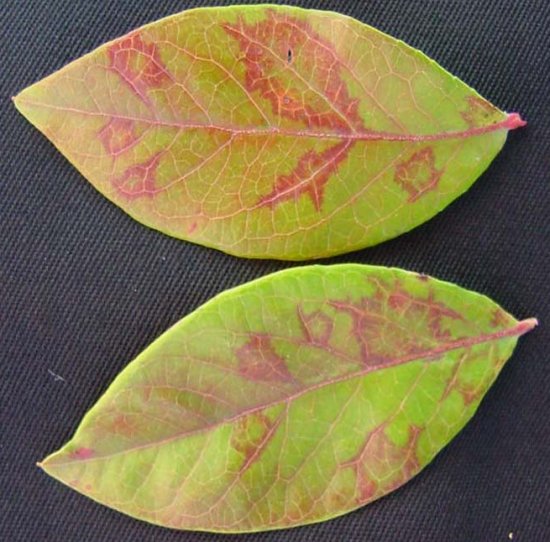
A virus with flexuous rod-shaped particles c. 690 nm in length by 14 nm in width (Martin & Bristow, 1988) (Fig. 1), which contains a single molecule of positive-sense ssRNA of 8514 bp and a single capsid protein of approximately 33,500 kDa (Cavileer et al., 1994). The virus is transmitted in a non-persistent manner by several aphid species and can be transmitted mechanically to Chenopodium quinoa and Nicotiana occidentalis with difficulty (Podleckis et al., 1986; Bernardy et al., 2004). The virus occurs in Vaccinium species in North America and Europe. In most cultivars of highbush blueberry (Vaccinium corymbosum) it causes various degrees of flower and leaf necrosis (Fig. 2, Fig. 3) but is symptomless or causes mild chlorosis in several cultivars (Fig. 4). It has been detected in several Vaccinium species native to the Cascade Mountains of western USA and caused symptoms similar to those observed in highbush blueberry. It has also been reported in cranberry (Vaccinium macrocarpon) and black huckleberry (Vaccinium membranaceum) where it is symptomless (Wegener et al., 2004; 2007).
Main Diseases
In most cultivars of highbush blueberry, infection with BlScV results in blighting of some of the flowers and leaves during the bloom period and in some cases flowers are retained into the following year and appear a silverish colour (Fig. 5). In other cultivars plants remained symptomless for at least 2-3 years after grafting (Bristow et al., 2000). Several cultivars develop an oakleaf pattern late in the season, but this is not a consistent symptom (Fig. 6). With strains of the virus from Washington and Oregon there were more cultivars that remained symptomless than was observed in New Jersey, where only the cultivar 'Jersey' was symptomless. In the initial tests there were several cultivars that did not become infected when grafted with BlScV (Bristow et al., 2000); these cultivars have since been regrafted and have become infected with the virus. There is no known source of resistance to BlScV in Vaccinium corymbosum. In cultivars that exhibit flower and leaf necrosis yield losses can exceed 80%, whereas in symptomless cultivars there is no observable yield loss due to infection with BlScV (Bristow et al., 2000).
Geographical Distribution
The virus has been found in the USA; both in the Northwest, in Oregon and Washington (Martin & Bristow, 1988) and the Northeast: New Jersey (Stretch, 1983), Connecticut and Massachusetts (DeMarsay et al., 2004). Also recorded in Canada, in British Columbia (Wegener et al., 2002) and in Europe; in the Netherlands (Martin, unpublished 2002) and Italy (Ciuffo et al., 2005). The recent finding of BlScV infecting black huckleberry asymptomatically in remote areas of British Columbia (Wegener et al., 2007) together with the diversity of strains reported from that region suggests the virus may have originated in native Vaccinium species in British Columbia.
Host Range and Symptomatology
The virus appears to be limited to the genus Vaccinium in natural settings,
with some species remaining symptomless. Due to the lack of symptoms in some hosts,
diagnosis using indicators is not reliable, with serological or nucleic acid-based methods
required. The experimental host range is limited.
- Diagnostic species
- Vaccinium corymbosum cvs. 'Berkeley', 'Pemberton', 'Bluetta', 'Earliblue', as well as many others flower and leaf necrosis develop at full-bloom (Fig. 2 and Fig. 3), the leaf necrosis persists throughout the season. The cultivar 'Jersey' has not shown these symptoms.
- Chenopodium quinoa - local lesions followed by a systemic mottling, the symptoms take up to three weeks to develop.
- Propagation species
- Vaccinium corymbosum, Chenopodium quinoa, Nicotiana occidentalis. In the latter host,symptoms are very subtle with a slight twisting of leaves on the stem, thus plants should be tested by ELISA or PCR to confirm infection prior to purification.
- Assay species
- Vaccinium corymbosum cvs. 'Berkeley' or 'Pemberton'. There is no local lesion host.
Strains
The NJ-2 strain of BlScV should be considered the type strain since it was the first sequenced (Cavileer et al., 1994). Two additional strains from British Columbia have been sequenced subsequently (Bernardy et al., 2004) and show considerable variation at the nucleotide level. Sequencing of the coat protein (CP) of multiple strains showed that there is considerable variation in this gene with 15-20% variation at the nucleotide level being very common with five additional strains suggested in British Columbia (Wegener et al., 2006). Surprisingly, serological variation of the CP has not been observed, as all isolates tested thus far, are detected by the antibodies produced when the virus was first described.
Transmission by Vectors
BlScV is transmitted inefficiently by the common blueberry aphid (Ericaphous fimbriata) and has been transmitted under laboratory conditions by several other species of aphids. All aphids tested that vectored the virus were inefficient vectors when transmitting from blueberry-to-blueberry. The common blueberry aphid is the aphid most commonly found colonizing blueberry in the Pacific Northwest (Oregon, Washington, British Columbia).
Transmission through Seed
No evidence for seed transmission has ever been found.
Serology
BlScV is a good immunogen and antiserum developed has been used for ELISA and western blots (Martin & Bristow, 1988; Martin et al., 1992). The pH of blueberry leaf sap after homogenizing in standard ELISA buffer is about 3.5 and it is necessary to use a buffer that will maintain a higher pH when grinding blueberry tissue for ELISA testing (Martin & Bristow, 1988; MacDonald et al., 1988).
Relationships
BlScV is a member of the genus Carlavirus in the family Flexiviridae (Mayo et al., 2005). The physical and chemical properties of BlScV are similar to those of other members of the genus Carlavirus and is related serologically or by sequence comparison to Carnation latent virus, Chrysanthemum virus B, Dandelion latent virus, Elderberry symptomless virus, Helenium virus S, Lily symptomless virus, Poplar mosaic virus, Potato virus M, Potato virus S and Red clover vein mosaic virus with a more distant relationship to Pea streak virus (Martin & Bristow, 1988; Cavileer et al., 1994).
Stability in Sap
No studies on its stability in sap have been reported.
Purification
Using method of
Martin & Bristow (1988).
Leaves of infected blueberry plants are homogenized in 0.1 M sodium borate, pH 8.2, containing 2% (w/v) PVP-44, 0.1 M EDTA, 0.5% (v/v) nicotine alkaloid and 0.1% (v/v) β-mercaptoethanol; using 5 ml per gram of tissue. The sap is expressed through cheesecloth and centrifuged at 10,000 g for 10 min. The supernatant is centrifuged at 110,000 g for 90 min. and the pellets resuspended in 0.01 M potassium phosphate, pH 7.5, containing 0.1% (v/v) PVP-44 and 1 mM EDTA. After low-speed centrifugation at 10,000 g for 10 min, the supernatant is centrifuged through a 30% sucrose cushion at 110,000 g for 2 hr. The pellets are resuspended in a phosphate buffer as above and CsCl is added to final density of 1.28 g/ml (3.86 g/10 of final volume). The samples are then centrifuged for 16 hr at 247,000 g and the opalescent band drawn off with a needle and syringe. After collection the band is diluted in 0.01 M phosphate buffer and centrifuged at 170,000 g for 1 hr, the final pellets are saved and resuspended in 0.01 M phosphate buffer, pH, 7.5.
Properties of Particles
The particles have a density of 1.28 g/ml based on banding in CsCl gradients (Martin & Bristow, 1988). Other physical properties have not been determined.
Particle Structure
The virus particles are slightly flexuous rods approximately 690 nm in length and 14 nm wide.
Particle Composition
Nucleic acid: Virus particles contain one species of positive-sense ssRNA. The complete nucleotide sequence has been determined for the NJ-2 isolate (Cavileer et al., 1994) and for BC-1 and BC-2 (Bernardy et al., 2004).
Protein: Polyacrylamide gel electrophoresis of the viral CP revealed a single polypeptide of approximately 35 kDa (Martin & Bristow, 1988), while the molecular weight of the putative CP calculated from published sequences ranges from 33.3 to 33.6 kDa (Cavilieer et al., 1994).
Genome Properties
The genome encodes for six proteins: ORF1 encodes a 223 kDa protein that is the putative replicase containing methyl transferase, NTP-binding site, helicase and polymerase domains and is followed by an intergenic region; ORF 2 -4 overlap and make up the triple gene block common to potexviruses, hordeiviruses, furoviruses and carlaviruses, ORF 2 codes for a 25 kDa protein that is a helicase homologue, ORF 3 codes for a 12 kDa basic protein and ORF 4 for a 7 kDa basic protein and is followed by a second intergenic region; ORF 5 codes for the 33 kDa CP; and a ORF 6 codes for a cysteine-rich protein that overlaps with the CP (Cavileer et al., 1994). The sequence of BC-1 and BC-2 shared nucleotide sequence identity of 83% and 77% with the NJ-2 sequence, respectively (Bernardy et al., 2004).
Relations with Cells and Tissues
BlScV is distributed in mesophyll cells, where the virus has been observed in large bundles (Martin & Bristow, 1988). Uneven distribution of virus in plants is quite common and it is recommended that when testing a plant at least three leaves each from a different cane be combined for the sample to be tested.
Ecology and Control
BlScV can cause significant damage in most blueberry cultivars tested. It has been observed in native species in British Columbia, Canada. It also infects cranberry without causing any symptoms and thus is not an important virus for cranberry growers. However, the blueberry aphid is able to transmit the virus from blueberry-to-cranberry and likely can transmit from cranberry-to-blueberry. Thus, cranberry plants can be an important means of long distance movement of BlScV into new areas. In Oregon, Washington and British Columbia approximately 1 in 6 cranberry bogs tested were found to have some vines infected with BlScV (Wegener et al., 2004). Cranberries are grown in areas that are diked and can be readily flooded. At harvest the fruit often is stripped from the plants after the "bog" has been flooded, the fruit floats and is collected onto trucks with an elevator. The close proximity of blueberry and cranberry production could explain the rapid spread of BlScV in blueberries in British Columbia, compared to that observed in Oregon and Washington, where cranberries are grown along the coast and blueberries inland.
The symptoms of BlScV infection can be confused easily with frost damage, Botrytis flower blight, Monilinia blossom blight, Pseudomonas infection or winter moth damage, stressing the need for confirmation of cause of symptoms before any control efforts are implemented.
Control of BlScV is primarily through the production of virus-tested nursery plants and should not be based on virus symptoms, since many cultivars do not express symptoms in nursery stock. Once a field has become infected a grower has two options to control BlScV. The first is removal of all plants in the field, which is recommended if the percentage of infected plants is high. If replanting with blueberries, it is important that the root system of the plants have been removed to prevent suckers growing from the infected roots system, which would provide a source of inoculum of BlScV. The second course of action is for cases where the incidence of infection is low, where a grower can combine aphid control with virus testing and removal. Aphid control should be implemented prior to removal of plants in order that aphids are not dispersed during the removal process. It is important to maintain aphid control for at least two years after the last infected plants have been removed to allow any recently infected plants to develop symptoms. It is also necessary to have virus detection based on ELISA (Martin & Bristow, 1988) or RT-PCR (Halpern & Hillman, 1996) to ensure that any strains that may not cause symptoms in the cultivars grown are also removed. This is necessary so that the grower is not restricted in cultivar selection in future plantings and to prevent movement of the virus into adjacent fields that may have other cultivars being grown. Testing should be repeated the year after removal of infected plants to identify plants that may have been infected shortly before testing, which could have resulted in false negatives if the virus titer was very low or the virus was minimally distributed in plant.
Figures
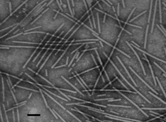
Particles of BlScV; bar represents 100 nm.

Whole blueberry plant infected with BlScV between healthy plants.
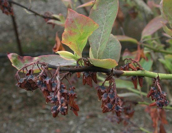
Close up of flower necrosis in 'Berkeley' blueberry.

Chlorosis observed on older leaves of some blueberry cultivars when infected with BlScV.
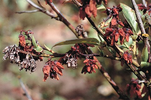
Symptomatic 'Berkeley' plant showing silverish flower clusters from previous year and freshly necrotic flower clusters from current year symptoms.
Oakleaf pattern observed in some blueberry cultivars late in the season when infected with BlScV.
References list for DPV: Blueberry scorch virus (415)
- Bernardy, Dubeau, Braun, Lowry & French, Canadian Journal of Plant Pathology 26: 405, 2004.
- Bristow, Martin & Windom, Phytopathology 90: 474, 2000.
- Cavileer, Halpern, Lawrence, Podleckis, Martin & Hillman, Journal of General Virology 75: 711, 1994.
- Ciuffo, Pettiti, Gallo, Masenga & Turina, Plant Pathology 54: 565, 2005.
- DeMarsay, Marucci, Hillman, Petersen, Oudemans, Marucci & Schloemann, Plant Disease 88: 572, 2004.
- Halpern & Hillman, Plant Disease 80: 219, 1996.
- MacDonald, Martin, Gillett & Ramsdell, Acta Horticulturae 236: 37, 1988.
- Martin & Bristow, Phytopathology 78: 1636, 1988.
- Martin, MacDonald & Podleckis, Acta Horticulturae 308: 131, 1992.
- Mayo, Maniloff, Desselberger, Ball & Fauquet, Virus Taxonomy: VIIIth Report of the International Committee on Taxonomy of Viruses, Elsevier Academic Press, 2005.
- Podleckis, Davis, Stretch & Schulze, Phytopathology 76: 1065, 1986.
- Raworth, Canadian Entomology136: 711, 2004.
- Stretch, Phytopathology 73: 375, 1983.
- Wegener, MacDonald, Sweeney, Joshi, Hudgins, French, Raworth & Martin, Canadian Journal of Plant Pathology 24: 91, 2002.
- Wegener, Martin, Bernardy, MacDonald & Punja, Canadian Journal of Plant Pathology 28: 250, 2006.
- Wegener, Punja & Martin, Plant Disease 88: 427, 2004.
- Wegener, Punja & Martin, Plant Disease 91: (in press), 2007.