Details of DPV and References
DPV NO: 44 June 1971
Family: Bromoviridae
Genus: Ilarvirus
Species: Tobacco streak virus | Acronym: TSV
There is a more recent description of this virus: DPV 307
Tobacco streak virus
R. W. Fulton Department of Plant Pathology, University of Wisconsin, Madison, Wisconsin, USA
Contents
- Introduction
- Main Diseases
- Geographical Distribution
- Host Range and Symptomatology
- Strains
- Transmission by Vectors
- Transmission through Seed
- Transmission by Grafting
- Transmission by Dodder
- Serology
- Nucleic Acid Hybridization
- Relationships
- Stability in Sap
- Purification
- Properties of Particles
- Particle Structure
- Particle Composition
- Properties of Infective Nucleic Acid
- Molecular Structure
- Genome Properties
- Satellite
- Relations with Cells and Tissues
- Ecology and Control
- Notes
- Acknowledgements
- Figures
- References
Introduction
-
Described by
Johnson (1936).
Selected synonyms
- Annulus orae
(Rev. appl. Mycol. 28: 514) - Nicotiana virus 8 (Rev. appl. Mycol. 36: 303)
- Nicotianavirus vulnerans (Rev. appl. Mycol. 36: 303)
- Tractus orae (Rev. appl. Mycol. 20: 85)
- Nicotianavirus vulnerans (Rev. appl. Mycol. 36: 303)
-
An isometric virus with particles 28 nm in diameter. It has a wide host range, but no known vector. It is readily transmitted by inoculation of sap, but is unstable in sap. The virus is wide-spread, but not common.
Main Diseases
Causes a systemic necrotic disease (‘necrose branca’) in tobacco, which recovers from symptoms; mottling or no symptoms in dahlia (Costa & Carvalho, 1961; Fulton, 1967; Brunt, 1968), mottling in cotton (Costa & Carvalho, 1961), Trifolium pratense and Melilotus alba; yellow ringspot and malformation of tomato (Martelli & Cirulli, 1969; Costa et al., 1961); stunting of asparagus (Brunt & Paludan, 1970); vein-yellowing in rose (Fulton, 1970b); and systemic necrotic symptoms in pea (Patino & Zaumeyer, 1959), potato (Costa, Carvalho & Deslandes, 1964) and soybean (Costa & Carvalho, 1961). It has also been isolated from grape (J. K. Uyemoto, pers. comm.), papaya (A. F. Ross, pers. comm.), black raspberry (R. H. Converse, pers. comm.), strawberry (Stace-Smith & Frazier, 1971), groundnut, velvet bean (Stizolobium deeringianum), Crotalaria and a number of weed species.
Geographical Distribution
Europe, North and South America, New Zealand.
Host Range and Symptomatology
The host range is wide; many species in 31 monocotyledonous and dicotyledonous families are susceptible.
Diagnostic species
- Nicotiana tabacumn
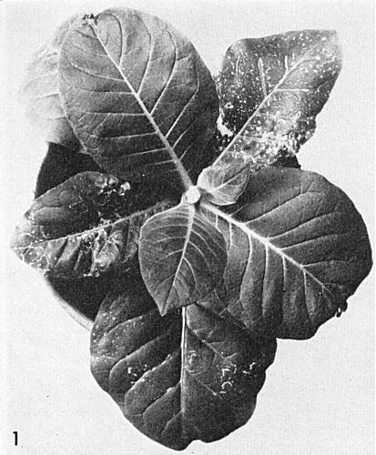
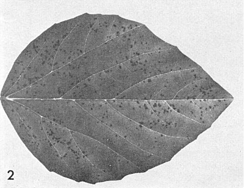
(tobacco). Local necrotic spots or rings, systemic necrotic lines and ‘oak-leaf’ patterns; plant recovers from necrotic symptoms, and leaves that expand several weeks after inoculation seem healthy (Fig. 1). With many strains, the ‘recovered’ leaves of Turkish tobacco have dentate rather than entire margins.- Cyamopsis tetragonoloba (guar). Small, dark local lesions
(Fig. 2).
- Vigna cylindrica (catjang). Local reddish necrotic lesions or rings, depending on the strain of virus (Fig. 3). Systemic vein-clearing or necrosis.
-
Propagation species
- Cultures may be maintained in tobacco or Vinca rosea. Tobacco, Nicotiana rustica, Datura stramonium and Vigna cylindrica are good sources of one or other strain of the virus for purification.
-
Assay species
- Cyamopsis tetragonoloba, Vigna cylindrica, Beta patellaris
and Phaseolus vulgaris cv. Manteiga.
Strains
The complex of variants isolated in Brazil is considered a distinct strain (Costa & Carvalho, 1961), differing from North American strains in host range and symptoms. Strains differing from the type in host range and properties have been obtained from bean with red node disease (Thomas & Zaumeyer, 1950) and pea. Variants are often distinguishable by the type of lesion they produce in Vigna cylindrica and by symptoms in other hosts.
Transmission by Vectors
No vectors have been reported.
Transmission through Seed
Reported for bean, Datura stramonium and Chenopodium quinoa, but not for other hosts tested (Brunt, 1969).
Transmission by Dodder
Transmitted by Cuscuta campestris (Fulton, 1948).
Serology
The virus is not strongly immunogenic, but 10-12 intramuscular injections at 3 or 4 day intervals of 1 mg of virus emulsified in Freund’s incomplete adjuvant gave antiserum titres of 1/1280 or more (by microprecipitin test). Precipitates in liquid tests are granular. The virus reacts well in agar double-diffusion tests.
Relationships
No cross-reactions with other viruses or serological differences between strains have been described. Strains of the virus reciprocally cross-protect. In the symptoms it induces, its properties, and the shape of its particles, the virus resembles the serologically unrelated Tulare apple mosaic virus.
Stability in Sap
Undiluted tobacco sap loses most of its infectivity within 5 min after extraction (Fulton, 1949), and all of its infectivity in less than 36 hr. Infectivity is lost more slowly in diluted extracts, and is stabilized by antioxidants, especially 2-mercaptoethanol. Thermal inactivation points between 53 and 64°C have been reported. Dilution end-points of extracts have been reported between 1/30 and 1/15,625. The virus remains infective for many years in diced tissue dried cold under vacuum and stored cold.
Purification
The following method is effective (Fulton, 1967). Homogenize heavily infected inoculated leaf tissue at the rate of 100 g in 150 ml of buffer (0.02 M phosphate, pH 8.0, containing 0.02 M 2-mercaptoethanol) plus 15 g Al2O3. After centrifuging 15-20 min at 1500 g, mix the supernatant liquid thoroughly with 80 ml hydrated calcium phosphate per 100 g tissue. Centrifuge at 1500 g again for 15-20 min. Sediment the virus from the supernatant liquid by centrifuging 3 hr at 78,000 g. Resuspend the pellets in 0.01 M disodium ethylenediamine-tetraacetate (EDTA), pH 6.0, adjust the pH to 5.0 with citric acid and remove the precipitate by centrifugation. Readjust the supernatant liquid, containing the virus, to pH 6.0, and concentrate the virus by high speed centrifugation. Purified virus is more stable in 0.01 M EDTA, pH 6.0, than in water or any of a number of buffers. Yields may be up to 45 mg virus per 100 g tissue. A procedure involving charcoal and freezing clarification, with differential centrifugation, has been used for the red node strain (Mink, Saksena & Silbernagel, 1966).
Properties of Particles
The virus has at least three kinds of nucleoprotein particles with sedimentation coefficients (s20, w) between about 90 and 113 S. The least rapidly sedimenting particles are non-infectious, the other two types are weakly infectious alone, but highly infectious when mixed. Adding the non-infectious particles to mixtures of the other two kinds further increases infectivity. When components from different strains are mixed the lesion type seems to be determined by the top or by the middle component (Fulton, 1970a). The proportion of the components varies with the host and with the method of tissue extraction (Lister & Bancroft, 1970).
Absorbance at 260 nm (1 mg/ml, 1 cm light path): 5.1.
A260/A280: c. 1.56.
Particle Structure
Particles are isometric, about 28 nm in diameter (Fig. 4).
Particle Composition
Not determined.
Relations with Cells and Tissues
No information.
Notes
Symptoms in experimental hosts may be confused with those caused by several other viruses. Identification should be made by cross-protection tests or by serology.
Figures
References list for DPV: Tobacco streak virus (44)
- Brunt, Pl. Path. 17: 119, 1968.
- Brunt, Rep. Glasshouse Crops Res. Inst. 1968: 104, 1969.
- Brunt & Paludan, Phytopath. Z. 69: 277, 1970.
- Costa & Carvalho, Phytopath. Z. 43: 113, 1961.
- Costa, Carvalho, Oliveira & Deslandes, Bragantia 20: cvii, 1961.
- Costa, Carvalho & Deslandes,Bragantia 23: i, 1964.
- Fulton, Phytopathology 38: 421, 1948.
- Fulton, Phytopathology 39: 231, 1949.
- Fulton, Virology 32: 153, 1967.
- Fulton, Virology 41: 288, 1970a.
- Fulton, Pl. Dis. Reptr 54: 949, 1970b.
- Johnson, Phytopathology 26: 285, 1936.
- Lister & Bancroft, Phytopathology 60: 689, 1970.
- Martelli & Cirulli, Phytopath. Mediterranea 8:154, 1969.
- Mink, Saksena & Silbernagel, Phytopathology 56: 645, 1966.
- Patino & Zaumeyer, Phytopathology 49: 43, 1959.
- Stace-Smith & Frazier, Phytopathology 41: (in press), 1971.
- Thomas & Zaumeyer, Phytopathology 40: 832, 1950.


