Details of DPV and References
DPV NO: 65 October 1971
Family: Bromoviridae
Genus: Cucumovirus
Species: Peanut stunt virus | Acronym: PSV
This virus is now regarded as a distinct strain of peanut stunt virus
Robinia mosaic virus
K. Schmelzer Institut für Phytopathologie Aschersleben der Deutschen Akademie der Landwirtschaftswissenschaften zu Berlin, Germany
Contents
- Introduction
- Main Diseases
- Geographical Distribution
- Host Range and Symptomatology
- Strains
- Transmission by Vectors
- Transmission through Seed
- Transmission by Grafting
- Transmission by Dodder
- Serology
- Nucleic Acid Hybridization
- Relationships
- Stability in Sap
- Purification
- Properties of Particles
- Particle Structure
- Particle Composition
- Properties of Infective Nucleic Acid
- Molecular Structure
- Genome Properties
- Satellite
- Relations with Cells and Tissues
- Ecology and Control
- Notes
- Acknowledgements
- Figures
- References
Introduction
-
Described by Atanasoff (1935) and
Schmelzer (1967a).
Selected synonyms
- Black locust true mosaic virus (Rev. appl. Mycol. 46: 1346)
- Echtes Robinienmosaik-Virus (Rev. appl. Mycol. 46: 1346)
-
A virus with isometric particles c. 40 nm in diameter with a wide host range. Transmissible by inoculation of sap, by aphids in the non-persistent manner and also by some Cuscuta species. The virus occurs commonly in south-eastern Europe and rarely in central Europe.
Main Diseases
Causes mosaic and leaf deformation of Robinia pseudo-acacia (Fig. 1).
Geographical Distribution
South-eastern and central Europe (Schmelzer, 1968).
Host Range and Symptomatology
Species of Robinia, especially R. pseudo-acacia, seem to be the only natural hosts, but about 75 plant species in 15 dicotyledonous families have been infected experimentally by sap inoculation (Schmelzer, 1967a), including the following:
-
Diagnostic species
- Chenopodium quinoa.
Chlorotic local lesions; systemic mosaic and leaf distortion. Some strains always infect systemically, others only in winter. - C. murale. Chlorotic dots and necrotic pin-point spots; not systemic.
- Lycopersicon esculentum (tomato). Systemic mosaic and leaf curling.
- Petunia hybrida. Necrotic local lesions (Fig. 5); most isolates not systemic.
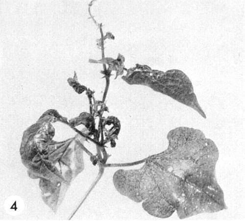
- Phaseolus vulgaris (French bean). Chlorotic or necrotic local lesions. All tested isolates infect at least some varieties systemically (Fig. 4), giving mosaic, necrosis, and deformation of tip leaves.
- Vigna sinensis (cowpea). Chlorotic or necrotic local lesions; systemic mosaic (Fig. 3).
- Lycopersicon esculentum (tomato). Systemic mosaic and leaf curling.
-
Propagation species
- Nicotiana glutinosa
(Fig. 6) is suitable both for maintaining cultures and as a source of virus for purification. - N. megalosiphon is also good for maintaining the virus.
- Useful amounts of the virus may be obtained from infected N. tabacum except in summer.
-
Assay species
- Chenopodium quinoa
is a good local lesion host; C. foetidum has been used in aphid transmission tests.
Strains
Not well studied. Isolates differ in virulence and host range. For example, strain Ro 25, isolated in Hungary (Schmelzer, 1967a), induces chlorotic local lesions as well as mosaic and leaf deformation in Chenopodium quinoa throughout the year, whereas less virulent isolates become systemic in this species only during the winter.
Transmission by Vectors
The aphids Myzus persicae and Aphis craccivora transmit the virus in the non-persistent manner. The transmission rate is rather low. The aphids acquire the virus within 10 min and there is no latent period. Aphis craccivora seems to be the most important vector in nature (Schmelzer & Milicic 1965).
Transmission through Seed
Not found.
Transmission by Dodder
Possible with Cuscuta californica, C. campestris and C. subinclusa. The virus infects the dodder (Schmelzer, 1967a).
Serology
The virus is moderately antigenic. Antisera with titres of 1/128 are readily obtained. The virus reacts well with specific antiserum in agar gel double diffusion tests, which may be done with extracts made by grinding infected Chenopodium quinoa leaves with an equal amount of 0.067 M phosphate buffer (pH 7) or 0.85% NaCl solution. Only one band of precipitate is formed (Schmelzer, 1967a).
Relationships
There seems to be no great serological difference between isolates that differ in their host plant reactions or between isolates obtained from different countries. There is complete cross-protection between different isolates of the virus.
Stability in Sap
Using Chenopodium quinoa as virus source and test plant, the thermal inactivation temperature lies between 66 and 72°C, the dilution end-point above 10-4, and infectivity is retained at 20-22°C for 5-10 days.
Purification
Homogenize infected tissue with an equal amount of 0.067 M phosphate buffer (pH 7.0) containing 0.3% (w/v) sodium sulphite and 0.3% (w/v) ascorbic acid. Shake the resulting extract thoroughly with twice its volume of ether and, after removing the ether and the coagulated plant material, with half a volume of carbon tetrachloride. Centrifuge at low speed and retain the aqueous phase. Concentrate the virus by one cycle of high and low speed centrifugation. Resuspend in neutral 0.067 M phosphate buffer.
Properties of Particles
Not investigated.
Particle Structure
Particles are isometric, c. 40 nm in diameter (Fig. 2).
Particle Composition
Unknown.
Relations with Cells and Tissues
All tissues seem susceptible with the possible exception of the meristematic regions.
Notes
Cucumber mosaic virus has properties similar to those of robinia mosaic virus, but seems to have smaller particles. Numerous plant species react in almost the same way to both viruses, but, unlike cucumber mosaic virus, robinia mosaic virus usually infects Petunia hybrida only locally and Vigna sinensis, Arachis hypogaea and most cultivars of Phaseolus vulgaris systemically. French bean cultivars that are not infected systemically show large necrotic local lesions, in contrast to the pinpoint lesions induced by cucumber mosaic virus.
Alfalfa mosaic virus also has a host range similar to that of robinia mosaic virus, but common strains infect Phaseolus vulgaris and Vigna sinensis only in inoculated leaves and all strains induce necrotic local or severe necrotic systemic reactions in tomato, which robinia mosaic virus never does. Also the bacilliform particles of alfalfa mosaic virus are unlike those of robinia mosaic virus.
Besides robinia mosaic virus, tomato black ring (both potato bouquet and beet ringspot serotypes) and strawberry latent ringspot viruses cause mosaic symptoms in Robinia (Schmelzer, 1967b). However, these viruses infect Chenopodium murale systemically, whereas robinia mosaic virus is confined to the inoculated leaves, and neither is transmitted by aphids or by the species of dodder that transmit robinia mosaic virus.
Figures

Naturally infected leaflets of Robinia pseudo-acacia.
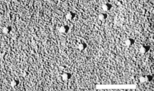
Virus particles from a purified preparation, shadowed with platinum and iridium. Bar represents 250 nm.

Systemically infected leaf of Vigna sinensis.
Systemic infection in Phaseolus vulgaris cv. Pinto.
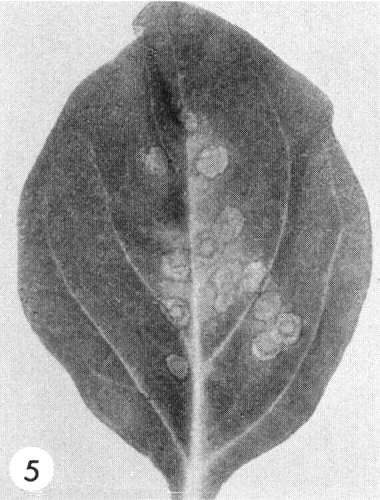
Local lesions in Petunia hybrida.

Systemic infection of Nicotiana glutinosa with strain Ro 25.
References list for DPV: Robinia mosaic virus (65)
- Atanasoff, Phytopath. Z. 8: 197, 1935.
- Schmelzer, Phytopath. Z. 58: 59, 1967a.
- Schmelzer, A XVII. Növényvédelmi tudományos Értekezlet 1: 139, 1967b.
- Schmelzer, Arch. Forstw. 17: 621, 1968.
- Schmelzer & Milicic, Acta bot. croat. 24: 189, 1965.