Details of DPV and References
DPV NO: 78 October 1971
Family: Potyviridae
Genus: Potyvirus
Species: Carnation vein mottle virus | Acronym: CVMoV
Carnation vein mottle virus
M. Hollings Glasshouse Crops Research Institute, Littlehampton, Sussex, England
Olwen M. Stone Glasshouse Crops Research Institute, Littlehampton, Sussex, England
Contents
- Introduction
- Main Diseases
- Geographical Distribution
- Host Range and Symptomatology
- Strains
- Transmission by Vectors
- Transmission through Seed
- Transmission by Grafting
- Transmission by Dodder
- Serology
- Nucleic Acid Hybridization
- Relationships
- Stability in Sap
- Purification
- Properties of Particles
- Particle Structure
- Particle Composition
- Properties of Infective Nucleic Acid
- Molecular Structure
- Genome Properties
- Satellite
- Relations with Cells and Tissues
- Ecology and Control
- Notes
- Acknowledgements
- Figures
- References
Introduction
- Described by Kassanis (1955).
- Synonyms
- None valid: Brierley & Smith (1957) proposed the name ‘carnation mosaic’ but this had previously been used for various other carnation viruses including carnation ringspot virus (Noordam, Thung & van der Want, 1951) and another isometric virus (Ames & Thornberry, 1952).
An RNA-containing virus with flexuous filamentous particles c. 790 nm long transmitted by the aphid Myzus persicae in the non-persistent manner, and by inoculation of sap. It causes disease in carnation and sweet william. Though uncommon in N.W. Europe, it is apparently more prevalent in USA and in some Mediterranean countries.
Main Diseases
In cultivated carnation (Dianthus caryophyllus) free from other viruses
it causes diffuse chlorotic spotting and mottling, with spots and flecks of darker
green on some of the veins of young leaves (Fig. 5). Older leaves are usually
symptomless. Symptoms may be very slight in some cultivars of the Sim group in winter,
but more conspicuous in others and in seedling clones (M. Hollings & O. M. Stone,
unpublished). Flower yield may be decreased and colour break and distortion may occur
(Fig. 3), especially in summer (Brierley, 1964a; Hakkaart, 1964). Symptoms are
intensified when the plants are also infected with carnation mottle virus (Fig. 2).
In sweet william (Dianthus barbatus), carnation vein mottle virus induces a conspicuous light and dark green leaf mottle, with flecks, rings and spots on the lateral veins (Fig. 1), and dwarfing and colour break in red or pink flowers (Kassanis, 1955).
Geographical Distribution
Apparently occurs wherever carnations are extensively grown; uncommon in glasshouse carnations in UK and countries of N.W. Europe where tests have been done, though commoner in S. Europe; widespread in Dianthus barbatus in gardens in UK (M. Hollings & O. M. Stone, unpublished). Occurs in USA (Brierley & Smith, 1957).
Host Range and Symptomatology
Apparently restricted to the Caryophyllaceae and allied families (Chenopodiaceae, Aizoaceae, Amaranthaceae, Portulacaceae, Polygonaceae and Plantaginaceae). Infects about 20 species in these families (M. Hollings & O. M. Stone, unpublished).
- Diagnostic species
- Chenopodium amaranticolor.
Local chlorotic and semi-necrotic lesions c. 1 mm diameter in 7-12 days; not systemic (Hollings, 1956). - C. quinoa. Local chlorotic spots in 7-10 days (dilute inocula often induce
no local lesions), systemic yellowish veinal flecks and spots in 2-4 weeks, with some
puckering, buckling and distortion (Fig. 4); more sensitive than C. amaranticolor
(Hollings & Stone, 1965).
- Dianthus barbatus. Selected clones that are resistant to infection with carnation mottle virus show systemic vein-clearing in the youngest leaves after c. 2 weeks, developing into a conspicuous leaf mottling as described above (Fig. 1).
- With Chenopodium species and most clones of D. barbatus, symptoms may be obscured if carnation mottle virus is also present.
- Dianthus barbatus. Selected clones that are resistant to infection with carnation mottle virus show systemic vein-clearing in the youngest leaves after c. 2 weeks, developing into a conspicuous leaf mottling as described above (Fig. 1).
- Propagation species
- Cultures are best maintained in D. barbatus clones resistant to infection
with carnation mottle virus.
- Assay species
- C. quinoa
and C. amaranticolor.
Strains
The few isolates tested produced similar symptoms in carnation and sweet william, but differed considerably in host range; some infected C. amaranticolor with difficulty (Hakkaart, 1964) or not at all (Weintraub & Ragetli, 1970). The type strain infected Gomphrena globosa and Amaranthus caudatus, causing systemic vein-mottle, but some other isolates do not infect these species. Minor differences in stability in vitro also occur.
Transmission by Vectors
Myzus persicae transmits the virus in a non-persistent manner (Kassanis, 1955). The virus can be acquired within 2 min and is transmitted by adult and larval instars (M. Hollings, unpublished). There is no evidence for virus multiplication within the vector. M. persicae f. dianthi (= M. polaris) was reported unable to transmit (Brierley & Smith, 1957).
Transmission through Seed
No seed-transmission detected in Dianthus barbatus or Chenopodium quinoa (M. Hollings & O. M. Stone, unpublished).
Transmission by Dodder
Cuscuta campestris did not transmit the virus from Chenopodium quinoa to C. quinoa (M. Hollings & O. M. Stone, unpublished).
Serology
The virus is a good immunogen. Rabbits given one intravenous injection, followed after 1 and 3 weeks by intramuscular injections (with Freund’s complete adjuvant) gave antisera with specific titres of up to 1/130,000 in precipitin tube tests with freshly purified antigen, and 1/8000-1/16,000 with antigen preparations stored for several days or more (M. Hollings & O. M. Stone, unpublished). Precipitates are flagellar. Unlike carnation latent virus, carnation vein mottle virus occurs in too low concentration in sap from Dianthus barbatus, D. caryophyllus and Chenopodium quinoa to enable reliable results to be obtained in tube precipitin tests. Intact virus particles do not diffuse in 0.8% agar gel and are not readily disrupted by ultrasound or by incorporating 1% (w/v) sodium dodecyl sulphate and 1% (w/v) 2-mercaptoethanol into the agar. However, virus protein obtained by disrupting purified virus with HCl reacted in gel-diffusion tests with antiserum to intact virus (M. Hollings & O. M. Stone, unpublished).
Relationships
Particle morphology and physico-chemical properties indicate that carnation vein mottle virus belongs to the potato virus Y group, and serological evidence supports this. Using purified virus preparations, a distant serological relationship was found to bean yellow mosaic, pea mosaic and freesia streak viruses, but not to 12 other viruses in the potato Y group (M. Hollings & O. M. Stone, unpublished).
Stability in Sap
Minor differences have been reported between isolates, but the stability of all lies within the normal range found with viruses of this morphology. Infectivity in sweet william sap survived heating (10 min) at 50 but not 55°C (Kassanis, 1955), or 60 but not 65°C (Brierley & Smith, 1957; Hollings, Stone & Thorne, 1971); it was lost after 2-10 days at c. 18°C or 22-28 days at c. 2°C. The dilution end-point in carnation sap lay between 10-2 and 10-5, in D. barbatus sap 10-3 to 10-4, and in C. quinoa sap 10-4 to 10-5. Lyophilized sap from D. barbatus stored at c. 18°C retained infectivity for 6 months (Hollings & Stone, 1970).
Purification
The virus tends to aggregate with host material and much can be lost in clarification; difficulties were reported by Kassanis (1955) and Weintraub & Ragetli (1970). Strong extracting buffers (0.5 or 0.2 M) are much better than weak ones (0.05 M), and Dianthus barbatus is better than C. quinoa or carnation as a virus source. The virus withstands n-butanol at 8.5% (v/v) for c. 1 hr (but not overnight). The following method gives good clarification and virus yield:
Homogenize young infected shoots of D. barbatus at laboratory temperature with 0.5 M phosphate buffer, pH 7.6, containing 0.1% thioglycollate (2-4 ml/g tissue) and allow the slurry to stand for 4-6 days at c. 4°C. Add n-butanol to the expressed juice to 8.5% of the total volume, and shake the mixture 45-60 min; separate the virus by one or more cycles of differential centrifugation, and resuspend the pellet in 0.02 vol 0.03 M phosphate buffer; remove insoluble matter by brief centrifugation at low speed (Hollings et al., 1971).
Preparations made by this method are purer, and have higher virus concentrations than those made by the methods of Wetter (1960) and Oertel (1969); yields may be up to 5 mg virus per 100 g leaf tissue extracted; preparations may have serological titres up to 1/2048. Further purification is possible by centrifugation in sucrose density gradients, in which the virus produces usually one, sometimes two, light-scattering zones. Virus recovered from the zones is then dialysed for 24 hr against 0.01 M phosphate buffer containing 0.07 M NaCl before pelleting.
Properties of Particles
Preparations contain only one sedimenting component; sedimentation coefficient (s20,w) at infinite dilution: about 144 S.
Absorbance at 260 nm (1 mg/ml, 1 cm light path): 2.1.
A260/A280: 1.15.
Particle Structure
Particles are elongated and flexuous c. 790 nm long and 12 nm wide, when stained in neutral phosphotungstate (Fig. 6) with catalase crystals as internal standard. When stained with uranyl formate, some internal particle structure is visible (Fig. 7). Particles differ greatly in length. The maximum length noted in thin sections was c. 750 nm (Weintraub & Ragetli, 1970).
Particle Composition
RNA: Probably single-stranded. Molar percentages of nucleotides: G21; A25; C25; U29 (M. Hollings & O. M. Stone, unpublished).
Protein: No information.
Relations with Cells and Tissues
Present in all parts of the plant including some meristematic tissues. Many kinds of cytoplasmic inclusions have been reported including dense bands, pinwheels, loops and circles. Cell organelles appear normal, except for the nuclei which are elongated and have abnormally electron-dense areas of chromatin. Nearly all virus-like particles in infected leaves were in a single thin layer between two membranes, just inside the tonoplast (Weintraub & Ragetli, 1970).
Notes
Carnation plants can be freed from the virus with some difficulty by heat treatment (Brierley, 1964b), but readily by meristem-tip culture (Stone, 1968).
Carnation vein mottle virus is readily distinguished from carnation latent virus by serology and electron microscopy, but most easily by inoculation of Dianthus barbatus, in which carnation latent virus causes no symptoms. The other carnation viruses (carnation mottle, carnation ringspot, carnation etched ring, Italian ringspot) all have isometric particles and cause distinctive symptoms in indicator plants.
Figures
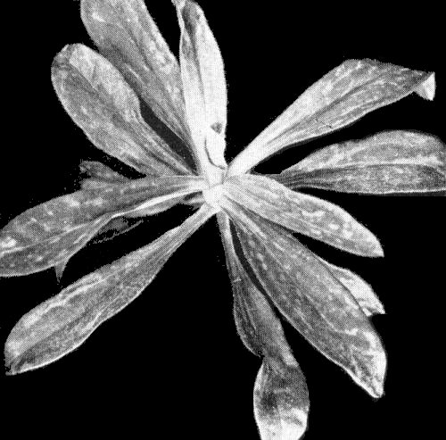
Dianthus barbarus, systemically infected.
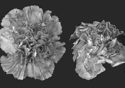
Flowers of carnation cv. La Reve; (right) infected with both carnation vein mottle and carnation mottle viruses (left) infected with carnation mottle virus only.
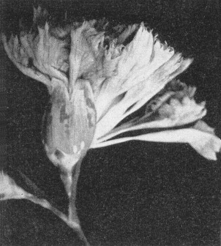
Infected flower of carnation seedling (Chabaud), showing calyx mottling.

Local lesions (left) and systemic yellowish flecks (right) in Chenopodium quinoa leaves.

Leaves of carnation cv. William Sim infected with carnation vein mottle virus only, showing faint darker green flecks.

Virus particles from a purified preparation stained with phosphotungstate. Bar represents 500 nm.

Particles in uranyl formate showing evidence of substructure. Bar represents 100 nm.
References list for DPV: Carnation vein mottle virus (78)
- Ames & Thornberry, Phytopathology 42: 289, 1952.
- Brierley, Pl. Dis. Reptr 48: 5, 1964a.
- Brierley, Pl. Dis. Reptr 48: 143, 1964b.
- Brierley & Smith, Phytopathology 47: 714, 1957.
- Hakkaart, Neth. J. Pl. Path. 70: 53, 1964.
- Hollings, Pl. Path. 5: 57, 1956.
- Hollings & Stone, Pl. Path. 14: 66, 1965.
- Hollings & Stone. Ann. appl. Biol. 65: 411, 1970.
- Hollings, Stone & Thorne, Rep. Glasshouse Crops Res. Inst. 1970: 148, 1971.
- Kassanis, Ann. appl. Biol. 43: 103, 1955.
- Noordam, Thung & van der Want, Tijdschr. PlZiekt. 57: 72, 1951.
- Oertel, Zentbl. Bakt. ParasitKde. Abt. II 123: 277, 1969.
- Stone, Ann. appl. Biol. 62: 119, 1968.
- Weintraub & Ragetli, Virology 40: 868, 1970.
- Wetter, Arch. Mikrobiol. 37: 278, 1960.