Details of DPV and References
DPV NO: 5 June 1970
Family: Bromoviridae
Genus: Ilarvirus
Species: Prunus necrotic ringspot virus | Acronym: PNRSV
Prunus necrotic ringspot virus
R. W. Fulton Department of Plant Pathology, University of Wisconsin, Madison, Wisconsin, USA
Contents
- Introduction
- Main Diseases
- Geographical Distribution
- Host Range and Symptomatology
- Strains
- Transmission by Vectors
- Transmission through Seed
- Transmission by Grafting
- Transmission by Dodder
- Serology
- Nucleic Acid Hybridization
- Relationships
- Stability in Sap
- Purification
- Properties of Particles
- Particle Structure
- Particle Composition
- Properties of Infective Nucleic Acid
- Molecular Structure
- Genome Properties
- Satellite
- Relations with Cells and Tissues
- Ecology and Control
- Notes
- Acknowledgements
- Figures
- References
Introduction
-
Described by Cochran & Hutchins (1941) and
Moore & Keitt (1944).
Selected synonyms
- Peach ringspot virus (Rev. appl. Mycol. 21: 85)
- Cherry (sour) necrotic ringspot virus (Moore & Keitt, 1944)
- Necrotic ringspot virus (Berkeley et al., 1951)
- Prunus ringspot virus (Rev. appl. Mycol. 39: 721)
- Cherry (sour) necrotic ringspot virus (Moore & Keitt, 1944)
-
An RNA-containing virus with isometric particles about 23 nm in diameter. Readily transmitted by inoculation with extracts of young leaves ground in buffer, but very labile. The virus has a fairly wide host range among dicotyledonous plants, no known insect vectors, but transmission by mites and nematodes has been reported. It is transmitted by pollen to seed and to pollinated plants.
Main Diseases
Causes necrotic ringspot in many species of Prunus (Fig. 5) (= Stecklenberg disease), often with subsequent recovery from symptoms. It causes sweet cherry rugose mosaic, almond calico (Nyland & Lowe, 1964), a mosaic disease of rose, and is common in hop (Bock, 1966; 1967), in which it is symptomless or associated with ring and band mosaic. Some strains and serotypes cause line pattern diseases of plum (Seneviratne & Posnette, 1970).
Geographical Distribution
Worldwide in temperate regions.
Host Range and Symptomatology
The host range is fairly wide; naturally and experimentally, the virus has infected species in 21 dicotyledonous families (Fulton, 1957a; Kirkpatrick, Lindner & Cheney, 1967).
-
Diagnostic species
- Cucumis sativus
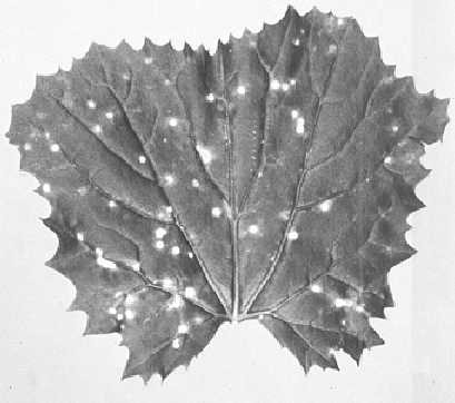
(cucumber). Prominent chlorotic primary lesions, systemic tip killing followed by extremely stunted and compact growth of axillary buds (Fig. 3). - Momordica balsamina. Necrotic primary lesions (Fig. 1),
occasionally systemic necrosis.
- Cyamopsis tetragonoloba. Large, dark local lesions (Fig. 2), systemic vein necrosis.
- Prunus serrulata ‘Shirofugen’ reacts to implanted virus-carrying buds with local necrosis and gumming.
- Cyamopsis tetragonoloba. Large, dark local lesions (Fig. 2), systemic vein necrosis.
-
Propagation species
- Prunus persica, P. mahaleb,
or Vinca rosea are suitable for maintaining cultures. As a source of virus for purification, cucumber cotyledons 3-5 days after inoculation are suitable; cherry petals also have been used (Tremaine & Willison, 1961).Assay species
- Momordica balsamina
is a good local lesion host; Cucumis sativus has also been used.
Strains
Many isolates differ slightly in herbaceous host range or symptomatology (Waterworth & Fulton, 1964). One common type infects Phaseolus vulgaris and Vigna sinensis systemically, other isolates do not. The recurrent strain causes ringspot symptoms each year on cherry; others produce symptoms only once. The Danish plum line pattern strain and the apple mosaic virus serotype cause line pattern symptoms in plum (Fulton, 1968).
Transmission by Vectors
No insect vector is known, in spite of extensive surveys (Swenson & Milbrath, 1964). The virus is pollen-borne in cherry and infects trees when they are pollinated with virus-carrying pollen (George & Davidson, 1964). Proeseler (1968) reported transmission by the mite Vasates fockeui; Fritzsche & Kegler (1968) reported transmission by the nematode Longidorus macrosoma.
Transmission through Seed
Transmitted in up to 70% of the seed of Prunus spp. (Megahed & Moore, 1967), although in a smaller proportion of seed of peach and some other species.
Transmission by Dodder
Not transmitted by Cuscuta campestris.
Serology
The virus is moderately immunogenic. Antiserum can be produced in rabbits by intramuscular injections of virus emulsified in Freund’s incomplete adjuvant. Injections are more effective at 3-4 day intervals than at longer intervals; intravenous injections are relatively ineffective. The virus reacts well in agar double diffusion tests and in liquid precipitin tests, in which precipitates are granular.
Relationships
Prunus necrotic ringspot virus is closely related serologically to Danish plum line pattern virus, but not to Cation’s Shiro plum line pattern virus. It is distantly serologically related to rose mosaic and apple mosaic viruses, which also cause line pattern of plum (Fulton, 1968). It may occur with, and somewhat resembles, prune dwarf virus, but there is no serological cross reaction between these two viruses.
Stability in Sap
In undiluted sap most infectivity is lost within a few minutes; in diluted sap maximum longevity is 9-18 hr. Infectivity is stabilized in extracts by 0.01 M Na-diethyldithiocarbamate but not by similar concentrations of cysteine hydrochloride or 2-mercaptoethanol (Fulton, 1957b). When infectivity is stabilized, thermal inactivation points (10 min) range from 55 to 62°C for different isolates (Waterworth & Fulton, 1964). Virus in tissue withstands rapid freezing to -78°C, but not slow freezing (Fulton, 1957b).
Purification
The following method is effective (Fulton, 1968). Homogenize heavily infected cucumber cotyledons cold in 1.5 ml buffer/g tissue. The buffer is 0.02 M phosphate, pH 8.0, and is 0.02 M with respect to 2-mercaptoethanol and Na-diethyldithiocarbamate. After low speed centrifugation, mix the supernatant liquid thoroughly with 0.8 volumes of hydrated calcium phosphate and again centrifuge at low speed for 10-20 min. Sediment the virus by centrifuging 3.5 hr at 78,000 g . Resuspend the pellets in phosphate buffer, bring to pH 4.8-5.0 with citric acid and remove the precipitate by centrifugation. Readjust the supernatant fluid, containing the virus, to pH 7 and concentrate the virus by high speed centrifugation. Density gradient centrifugation (van Regenmortel & Engelbrecht, 1963), density gradient electrophoresis (van Regenmortel, 1964) or precipitation of host protein by anti-host serum (Fulton, 1968) have been used for further purification.
Properties of Particles
The virus has two kinds of particles, with sedimentation coefficients (s20, w) variously reported as 79-97 S and 107-119 S. Both particle types are reported to be infective.
Molecular weight: 5.2-7.3 x 10⁶.
A260/A280: c. 1.56.
Particle Structure
Particles are isometric, 22-23 nm in diameter (Fig. 4). They disintegrate readily in phosphotungstate unless fixed first in 1% glutaraldehyde.
Particle Composition
RNA: About 16% of particle weight. Molar percentages of the nucleotides: G27; A25; C21; U27 (Barnett & Fulton, 1969).
Protein: Subunits have a molecular weight of about 2.5 x 10⁴ and contain about 196 amino acid residues (Barnett & Fulton, 1969).
Relations with Cells and Tissues
No information.
Notes
Prune dwarf virus causes shock symptoms in sour cherry resembling those caused by prunus necrotic ringspot virus but they are usually milder and involve only leaves that are unfolded and partially expanded, whereas those caused by prunus necrotic ringspot virus appear in small leaves before they unfold.
Figures
References list for DPV: Prunus necrotic ringspot virus (5)
- Barnett & Fulton, Virology 39: 556, 1969.
- Berkeley, Cation, Hildebrand, Keitt & Moore, U.S.D.A. Handbook 10: 164, 1951.
- Bock, Ann. appl. Biol. 57: 131, 1966.
- Bock, Ann. appl. Biol. 59: 437, 1967.
- Cochran & Hutchins, Phytopathology 31: 860, 1941.
- George & Davidson, Can. J. Pl. Sci. 44: 383, 1964.
- Fritzsche & Kegler, Tag. Ber. dt. Akad. Landw. Wiss. Berl. 97: 289, 1968.
- Fulton, Phytopathology 47: 215, 1957a.
- Fulton, Phytopathology 47: 683, 1957b.
- Fulton, Phytopathology 58: 635, 1968.
- Kirkpatrick, Lindner & Cheney, Pl. Dis. Reptr 51: 786, 1967.
- Megahed & Moore, Phytopathology 57: 821, 1967.
- Moore & Keitt, Phytopathology 34: 1009, 1944.
- Nyland & Lowe, Phytopathology 54: 1435, 1964.
- Proeseler, Phytopath. Z. 63: 1, 1968.
- Seneviratne & Posnette, Ann. appl. Biol. 65: 115, 1970.
- Swenson & Milbrath, Phytopathology 54: 399, 1964.
- Tremaine & Willison, Can. J. Bot. 39: 1387, 1961.
- van Regenmortel, Virology 23: 495, 1964.
- van Regenmortel & Engelbrecht, S. Afr. J. agric. Sci. 6: 505, 1963.
- Waterworth & Fulton, Phytopathology 54: 1155, 1964.


