Details of DPV and References
DPV NO: 53 June 1971
Family: Potyviridae
Genus: Potyvirus
Species: Beet mosaic virus | Acronym: BtMV
Beet mosaic virus
G. E. Russell Plant Breeding Institute, Trumpington, Cambridge, England
Contents
- Introduction
- Main Diseases
- Geographical Distribution
- Host Range and Symptomatology
- Strains
- Transmission by Vectors
- Transmission through Seed
- Transmission by Grafting
- Transmission by Dodder
- Serology
- Nucleic Acid Hybridization
- Relationships
- Stability in Sap
- Purification
- Properties of Particles
- Particle Structure
- Particle Composition
- Properties of Infective Nucleic Acid
- Molecular Structure
- Genome Properties
- Satellite
- Relations with Cells and Tissues
- Ecology and Control
- Notes
- Acknowledgements
- Figures
- References
Introduction
- Described by Lind (1915), Robbins (1921) and Smith (1957).
- Selected synonyms
- Beta
virus 2 (Rev. appl. Mycol. 17: 52) - Selected synonyms
- Marmor betae (Rev. appl. Mycol. 19: 229)
- A virus with flexuous filamentous particles c. 730 nm long and 13 nm wide. Transmitted by many species of aphids in the non-persistent manner and fairly easily by sap inoculation. Host range moderately wide. Widely distributed throughout the beet-growing areas of the world.
Main Diseases
Causes a mosaic disease in Beta vulgaris (sugar beet, red beet, spinach beet, etc.) and in Spinacea oleracea (spinach).
Geographical Distribution
World-wide in major beet-growing areas, especially in temperate regions.
Host Range and Symptomatology
Host range is moderately wide, mainly in the Chenopodiaceae, Solanaceae and Leguminosae; species in about 10 dicotyledonous families have been infected experimentally (Bennett, 1949). Transmitted readily by aphids or by sap inoculation to the following species:
- Diagnostic species
- Beta vulgaris
(sugar beet). Young leaves often show vein-clearing (Fig. 1). Older leaves show a pronounced light and dark green mottle and are often puckered (Fig. 3). Infected plants are sometimes stunted but severe distortion of the leaves is uncommon. - Spinacea oleracea (spinach). Numerous, small yellow flecks, often
coalescing to form large chlorotic areas, appear on the youngest leaves. Older
leaves become progressively chlorotic and necrotic, and infected plants are
usually stunted.
- Propagation species
- Sugar beet is ideal for maintaining cultures.
- Assay species
- Sugar beet and spinach are good hosts for testing aphids and for whole-plant assay using sap inoculation. Beta patellaris is a useful local lesion host, developing a few large necrotic lesions (Fig. 2). Amaranthus retroflexus and A. caudatus also give local lesions (Bennett, 1949). Many chlorotic local lesions are produced in Chenopodium quinoa (Fig. 4) and in Gomphrena globosa.
Strains
Many minor variants, differing in virulence towards sugar beet, have been described (Bennett, 1964). Beet water mottle virus (Watson, 1958) is probably a variant.
Transmission by Vectors
Transmissible by more than 28 aphid species (Kennedy, Day & Eastop, 1962) but Myzus persicae and Aphis fabae are the principal vectors in the field. Transmission is of the non-persistent type (Watson, 1946; Sylvester, 1949) and is improved by starving aphids for 2-5 min before acquisition feeds. Acquisition and inoculation thresholds are 6-10 sec; no latent period. Persistence of the virus in the vector depends on the species of aphid (Sylvester, 1952) but is probably less than 1 hr in apterae. All instars transmit but alatae transmit more efficiently than apterae and retain virus for up to 4 hr (Cockbain, Gibbs & Heathcote, 1963). The virus is not transmitted to progeny of vectors and vectors do not retain virus after moulting.
Transmission through Seed
Probably not seed-transmitted.
Transmission by Dodder
Three species of Cuscuta did not transmit (Bennett, 1944).
Serology
The virus is only moderately immunogenic (Bercks, 1960); antisera with titres greater than 1/1000 are difficult to obtain. Tube and droplet precipitin tests with clarified sap or partially purified virus preparations have been used.
Relationships
A member of the potato virus Y group of viruses (Brandes & Bercks, 1965) and serologically distantly related to bean yellow mosaic virus and potato virus Y (Bercks, 1960).
Stability in Sap
In beet sap, thermal inactivation point (10 min) is between 55 and 60°C, dilution end-point is up to 1/4000 and infectivity is retained for 24-48 hr at 20°C (Bennett, 1964). In beet leaves, infectivity survived 1 year at -20°C (Yamaguchi, 1964).
Purification
The virus seems rather unstable in vitro; partially purified preparations have been obtained using the following method (Chod & Polák, 1969). Extract frozen beet leaves in phosphate buffer (pH 7.8) containing 0.01 M veronal, 0.01 M cysteine and 0.007 M ethylenediamine-tetraacetate. Centrifuge at low speed. Precipitate the virus from the supernatant fluid by adding a 40% aqueous solution of polyethylene glycol (PEG) to reach a final PEG concentration of about 10%, and allowing the mixture to stand for 30 min. Sediment and clarify by differential centrifugation, resuspending the pellets obtained at high speed in 0.01 M phosphate buffer.
Properties of Particles
Extraction of leaves of infected sugar beet or Stellaria media with water-saturated phenol yielded preparations which infected Chenopodium amaranticolor (Beiss, 1963).
Particle Structure
Flexuous, filamentous particles (Fig. 5) about 730 nm long and 13 nm in diameter (Zimmer & Brandes, 1956).
Relations with Cells and Tissues
In beet, vesicular X-bodies are produced in the cytoplasm, and crystalline inclusions occur in the chloroplasts (Fujisawa, Matsui & Yamaguchi, 1967); nucleoli are enlarged and distorted in beet (Bos, 1969) and in Gomphrena globosa (Martelli & Russo, 1969); cytoplasm contains inclusions which appear in section as ‘pinwheels’ and bundles and are digestible by pepsin but not by ribonuclease (Hoefert, 1969).
Notes
Beet mosaic virus is usually of little economic importance in sugar beet or spinach; its effects on yield of sugar beet are very small (Wiesner, 1959). The symptoms it causes in sugar beet might be confused with the yellow mosaic and blotching caused by tomato black ring and tobacco rattle viruses (Gibbs & Harrison, 1964), though these two viruses are readily distinguished from beet mosaic virus by their symptoms in other hosts, the morphology of their particles, and by having nematode vectors. Among other viruses occurring in sugar beet and spinach, cucumber mosaic and beet ring mottle viruses (Duffus & Costa, 1963) cause mottling, pronounced chlorosis, narrowing and distortion of the leaves, and severely stunt the plants; beet yellows, beet mild yellowing and beet western yellows viruses all cause chlorosis of the older leaves but not mottling or chlorotic flecking; and beet curly top virus induces leaf-curling, wart-like protuberances on the leaves, and stunting of the plant.
Acknowledgements
Photographs: courtesy of Plant Breeding Institute, Cambridge; Fig. 5, courtesy of Rothamsted Experimental Station.
Figures

Young sugar beet leaves, (left) systemically infected, showing vein-clearing and incipient mottle symptoms; (right) virus-free.

Beta patellaris leaf showing brown necrotic local lesions.

Mature leaf from systemically infected sugar beet plant showing light green and dark green mottle symptoms.
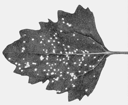
Chenopodium quinoa leaf showing chlorotic local lesions.

Electron micrograph of particle mounted in phosphotungstate. Bar represents 100 nm.
References list for DPV: Beet mosaic virus (53)
- Beiss, Naturwissenschaften 50: 675, 1963.
- Bennett, Phytopathology 34: 905, 1944.
- Bennett, Phytopathology 39: 669, 1949.
- Bennett, J. Am. Soc. Sug. Beet Technol. 13: 27, 1964.
- Bercks, Virology 12: 311, 1960.
- Bos, Neth. J. Pl. Path. 75: 137, 1969.
- Brandes & Bercks, Adv. Virus Res. 11: 1, 1965.
- Chod & Polák, Biologia Pl. 11: 324, 1969.
- Cockbain, Gibbs & Heathcote, Ann. appl. Biol. 52: 133, 1963.
- Duffus & Costa, Phytopathology 53: 1422, 1963.
- Fujisawa, Matsui & Yamaguchi, Phytopathology 57: 210, 1967.
- Gibbs & Harrison, Pl. Path. 13: 144, 1964.
- Hoefert, Virology 37: 498, 1969.
- Kennedy, Day & Eastop, A conspectus of aphids as vectors of plant viruses, London, Commonwealth Institute of Entomology, 1962.
- Lind, Tidsskr. PlAvl 22: 444, 1915.
- Martelli & Russo, Virology 38: 297, 1969.
- Robbins, Phytopathology 11: 349, 1921.
- Smith, Textbook of plant viruses, London, Churchill, 1957.
- Sylvester, Phytopathology 39: 417, 1949.
- Sylvester, Phytopathology 42: 252, 1952.
- Watson, Proc. R. Soc., B 133: 200, 1946.
- Watson, Rep. Rothamsted exp. Stn, 1957: 113, 1958.
- Wiesner, Zucker 12: 266, 1959.
- Yamaguchi, Ann. phytopath. Soc. Japan 29: 52, 1964.
- Zimmer & Brandes, Phytopath. Z. 26: 439, 1956.