Details of DPV and References
DPV NO: 286 July 1984
Family: Luteoviridae
Genus: Luteovirus
Species: Bean leafroll virus | Acronym: BLRV
Bean leaf roll virus
J. W. Ashby Plant Diseases Division, DSIR, Private Bag, Christchurch, New Zealand
Contents
- Introduction
- Main Diseases
- Geographical Distribution
- Host Range and Symptomatology
- Strains
- Transmission by Vectors
- Transmission through Seed
- Transmission by Grafting
- Transmission by Dodder
- Serology
- Nucleic Acid Hybridization
- Relationships
- Stability in Sap
- Purification
- Properties of Particles
- Particle Structure
- Particle Composition
- Properties of Infective Nucleic Acid
- Molecular Structure
- Genome Properties
- Satellite
- Relations with Cells and Tissues
- Ecology and Control
- Notes
- Acknowledgements
- Figures
- References
Introduction
First described by Quantz & Völk (1954) as ‘Blattrollkrankheit der Ackerbohne und Erbse’ (leaf roll disease of Vicia beans and peas). Virus characterised by Ashby & Huttinga (1979).
- Selected synonyms
- Pea leaf roll virus (Rev. appl. Mycol. 34: 272)
- Pea top yellows virus (Rev. appl. Mycol. 35: 144)
- Pea tip yellowing virus (Rev. appl. Mycol. 34: 505)
- Legume yellows virus (Rev. appl. Mycol. 59, 1500)
- Pea top yellows virus (Rev. appl. Mycol. 35: 144)
A virus with RNA-containing particles about 27 nm in diameter sedimenting as a single component. Hosts mainly in the Leguminosae. Transmitted by aphids in a persistent manner but not by inoculation of sap. Common in Europe, Middle East, India and USA.
Main Diseases
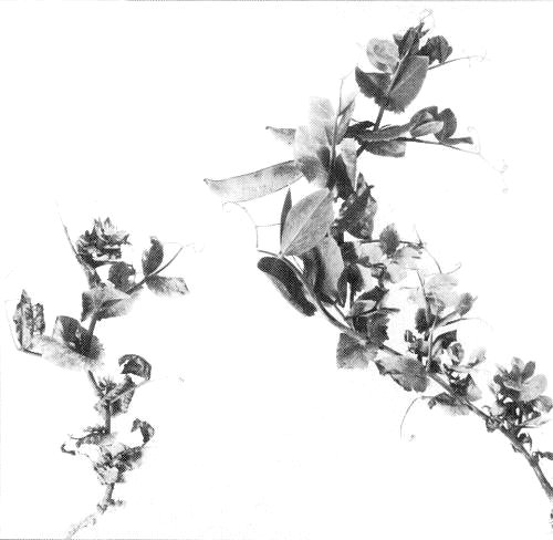
In broad bean (Vicia faba) the virus causes upward leaf-rolling accompanied by interveinal yellowing of older leaves (Fig. 2) and a decrease in number of pods (Quantz & Völk, 1954). In susceptible varieties of pea (Pisum sativum) it causes general stunting, chlorosis of the youngest leaves and sometimes downward leaf-rolling (Fig. 1, Fig. 4) (Quantz & Völk, 1954). The virus also causes stunting and yellowing of French bean (Phaseolus vulgaris), chickpea (Cicer arietinum), cowpea (Vigna unguiculata) and lentil (Lens esculenta) (Kaiser, 1972; Bos, 1982). Infection in the major perennial hosts, lucerne (alfalfa; (Medicago sativa)) and white clover (Trifolium repens), is usually symptomless. In lucerne, the virus has been associated with a vein-yellowing symptom (Van der Want & Bos, 1959) (Fig. 3) but this symptom is considered by Cockbain & Gibbs (1973) to be caused by another aphid-transmitted agent which requires the presence of BLRV for transmission.
Geographical Distribution
Bean leaf roll virus is common in Europe (Quantz & Völk , 1954; De Fluiter & Hubbeling, 1955 ; Roland, 1955; Cockbain & Gibbs, 1973), the Middle East (Kaiser, 1972), North Africa and India (Bos, 1982). The virus was tentatively identified from lucerne in Michigan (Thottappilly et al., 1977) and has recently been identified from pea crops in Pacific Northwest USA (Hampton, 1983). Diseases reported to have been caused by this virus in New Zealand (Wilson & Close, 1973) are now considered to have been caused by beet western yellows virus (Johnstone et al., 1984).
Host Range and Symptomatology
Not transmissible by mechanical inoculation of sap. Experimentally the virus was transmitted by means of aphids to 25 of 43 species of Leguminosae tested (Cockbain & Gibbs, 1973; Johnstone et al., 1984) and to three of 17 non-legumes tested: Erodium cicutarium, E. moschatum (Geraniaceae) and Montia perfoliata (Portulacaceae) (Johnstone et al., 1984). Thottappilly (1968) transmitted the virus to 65 species of Vicia, Pisum, Medicago, Trifolium, Lathyrus and Trigonella, genera on which the pea aphid occurs naturally. Development of symptoms in most species is greatly enhanced by the use of supplementary lighting and temperatures between 15 and 20°C (Johnstone et al., 1984).
- Diagnostic species
- Pisum sativum (pea). Symptoms are as described above,
but different pea cultivars produce a wide range of responses.
The following have proved useful (Drijfhout, 1968; Thottappilly
et al., 1977; Johnstone et al., 1984; R. O. Hampton,
personal communication).
-
Sensitive: Onyx (Fig. 4), Laxton’s Superb, Sounder, Charger.
Intermediate: Coquette, Salvo.
Tolerant: Ivy, Sybo, Sparkle.
Immune: Cobri, Lord Chancellor.- Trifolium subterraneum (subterranean clover) cv. Bacchus Marsh and Trifolium incarnatum (crimson clover) cv. Dixie. Mild interveinal yellowing followed by intense reddening, beginning at the margins of older leaves (Fig. 5).
- Beta vulgaris (beet) and Capsella bursa-pastoris (shepherd’s purse). Immune to infection (Johnstone et al., 1984).
- Trifolium subterraneum (subterranean clover) cv. Bacchus Marsh and Trifolium incarnatum (crimson clover) cv. Dixie. Mild interveinal yellowing followed by intense reddening, beginning at the margins of older leaves (Fig. 5).
- Propagation species
- Pisum sativum cvs Onyx, Eurofin, Charger and Gallaxie are
suitable for maintaining cultures and as a source of virus for
purification.
- Assay species
- Small seedlings of Trifolium subterraneum cv. Bacchus Marsh or T. incarnatum cv. Dixie are suitable for assaying transmission by aphid vectors.
Strains
Differences in aphid-transmissibility between two isolates of the virus have been reported (Cockbain & Costa, 1973).
Transmission by Vectors
The principal vector of bean leaf roll virus is the pea aphid, Acyrthosiphon pisum. Myzus persicae is a less efficient vector (Tinsley, 1959; Thottappilly, 1969; Cockbain & Costa, 1973). Macrosiphum euphorbiae is also a vector (Quantz & Völk, 1954). A single transmission by Megoura viciae was recorded by Quantz & Völk (1954) but Cockbain & Costa (1973) found this species to be a non-vector. Aphis fabae is a non-vector of European isolates (De Fluiter & Hubbeling, 1955; Thottappilly, 1969; Cockbain & Costa, 1973) but is reported to transmit the virus in Iran, as are Aphis cracccivora, Aphis gossypii, Acyrthosiphon pisum, Acyrthosiphon sesbaniae and Myzus persicae (Kaiser, 1972).
Transmission of the virus by Acyrthosiphon pisum at 18-24°C requires a minimum acquisition access period of 2 h, a median latent period (Sylvester, 1965) of 105 h and a minimum inoculation access period of 15 min. The virus persists in A. pisum through ecdysis and for up to 16 days, and probably does not multiply in the vector (Cockbain & Costa, 1973). Transmission of American isolates by A. pisum required a 2-4 h acquisition access period and a 2-4 h inoculation access period (R. O. Hampton, personal communication).
Transmission through Seed
The virus was not transmitted in seed from several virus-infected legumes (Kaiser, 1972).
Transmission by Dodder
Not transmitted by dodder (Cuscuta spp.) (Kaiser, 1972).
Serology
The virus is a good immunogen. Rabbit antisera with titres of 1/512 to 1/1024 in Ouchterlony double diffusion tests were obtained (Ashby & Huttinga, 1979). Antisera have also been shown to react with the virus and/or with related luteoviruses in immune density gradients (Ashby & Huttinga, 1979), immunosorbent electron microscopy (ISEM) (Roberts, Tamada & Harrison, 1980) and in enzyme linked immunosorbent assay (ELISA) (Hampton, 1983). Virus was easily detected by ELISA in several hosts including lucerne in which concentrations are very low (R. O. Hampton, personal communication).
Relationships
In gel diffusion tests (Ashby & Huttinga, 1979) bean leaf roll virus was found to react strongly (titre 1/128) with antiserum to soybean dwarf virus (homologous titre 1/1024) and weakly (1/8) with antiserum to beet western yellows virus (homologous titre 1/625 in infectivity neutralisation assay). Reactions with these antisera were also found in immune density gradients (Ashby & Huttinga, 1979). Antiserum to bean leaf roll virus (titre 1/512) reacted with purified subterranean clover red leaf virus, which is closely related to soybean dwarf virus, at a titre of 1/16 in gel diffusion tests (Ashby & Kyriakou, 1983). Antiserum to bean leaf roll virus also reacted weakly with purified potato leafroll virus (Roberts et al., 1980) and with purified carrot red leaf virus (Waterhouse & Murant, 1981). The luteoviruses appear to comprise a continuum of serologically related members.
In comparative host range studies (Johnstone et al.,
1984) bean leaf roll virus was shown to be closely similar to a
virus isolated from lucerne in Michigan (Thottappilly et al.,
1977) and a little less similar to the legume yellows virus reported
by Duffus (1979); soybean dwarf virus was not included in these
studies. Using ELISA, Hampton (1983) showed legume yellows, the
lucerne virus from Michigan and bean leaf roll virus from peas
and lucerne in Oregon all to react equally with antiserum
prepared to a Dutch isolate of bean leaf roll virus.
Stability in Sap
Acyrthosiphon pisum was unable to transmit the virus after feeding through Parafilm membranes on crude sap from infected plants, presumably because the concentration of virus in such extracts is too low. The aphid could however transmit virus after feeding in the same manner on purified virus preparations. Such preparations still contained transmissible virus after more than 10 days at room temperature (J. W. Ashby, unpublished data).
Purification
(Ashby & Huttinga, 1979). Grow infected pea plants at 15-20°C and harvest 2 wk after inoculation. Homogenise in 4 ml/g of cold 0.1 M phosphate buffer, pH 7.4, containing 0. 1% 2-mercaptoethanol and 10 mM EDTA using a Waring blender. Disintegrate further at higher speed (e.g. in Virtis 45 homogeniser) for 2 min. Centrifuge crude extract at 10,400 g for 10 min and add polyethylene glycol (to 8% w/v) and NaCl (to 3% w/v) to the supernatant fluid. Stir at 4°C for 1 h and centrifuge at 16,300 g for 15 min. Resuspend precipitate in extraction buffer and emulsify with 0.25 vol. of a 1:1 mixture of chloroform and n-butanol. After 1 h, separate phases by centrifugation. Recover virus from the aqueous phase by two cycles of differential centrifugation, resuspending the pellets in 50 mM phosphate buffer, pH 6.5, containing 1 mM EDTA. The virus may be further purified by centrifugation through a 10-40% linear sucrose density gradient (76,000 g, 3.5 h) followed by isopycnic density gradient centrifugation in caesium sulphate. Higher yields of virus (up to 668 µg/kg) were obtained from root tissue than from shoots (Ashby & Huttinga, 1979).
In recent years high yields of many luteoviruses have been
obtained by enzyme extraction methods (Takanami & Kubo, 1979).
Such methods are in general more efficient than that described above
and have proved effective for purification of bean leaf roll virus
(R. O. Hampton, personal communication).
Properties of Particles
In sucrose density gradients the particles sediment as a single component (Ashby & Huttinga, 1979).
Buoyant density in Cs2SO4: 1 .32 g/cm3.
A260/A280 : 1 .83 (not corrected for light-scattering).
Particle Structure
Particles are isometric, about 27 nm diameter, in preparations negatively stained with uranyl acetate (Fig. 6) (Ashby & Huttinga, 1979).
Particle Composition
Nucleic acid: Single-stranded RNA with estimated M. Wt c. 2.4 x 10⁶ under non-denaturing conditions.
Protein: In SDS/polyacrylamide gel electrophoresis, a single polypeptide was detected, with estimated M. Wt 32,500 (Ashby & Huttinga, 1979). This seems unusually high and the value of 23,000 obtained by R. O. Hampton (personal communication) is more typical for a luteovirus.
Relations with Cells and Tissues
Virus-like particles were observed in necrotic phloem sieve-tube cells of virus-infected broad bean and crimson clover. The particles were scattered singly or in masses throughout the lumen of infected cells and were interspersed with remnants of cell organelles (Thottappilly et al., 1977).
Notes
Confusion over the proper name for bean leaf roll virus and the disease caused by it were discussed by Bos (1964) who suggested that preference be given to the name pea leaf roll virus. The first description of the disease ‘Blattrollkrankheit der Ackerbohne’ was by Böning (1927) and the viral nature of the disease in Vicia-beans and peas was demonstrated by Quantz & Völk (1954). The name leaf roll is descriptive of the symptoms caused in beans whereas in peas the main symptom is apical yellowing. Thus on the basis of priority and descriptiveness bean leaf roll is the most appropriate name for the virus.
Several luteoviruses of legumes resemble bean leaf roll virus
and apparent differences between some of them were described by
Tamada & Kojima (1977). There is increasing evidence that
one of those described, subterranean clover stunt virus, may not
be a member of the luteovirus group (G. R. Johnstone, personal
communication). A tentative proposal for grouping the luteoviruses
that resemble bean leaf roll virus is given in Table 1. A list of
key hosts and of procedures for studying host range and vector
specificity is given by Johnstone et al. (1984). Although
imprecise, these methods permit workers who do not have access to
such techniques as ISEM and ELISA to make a tentative
identification of luteoviruses isolated from leguminous hosts.
Among such viruses, beet western yellows virus is the one most
likely to be confused with bean leaf roll virus; however, although
beet western yellows virus has a wide host range in the Leguminosae,
it also infects non-legumes such as Capsella bursa-pastoris, Beta
vulgaris and Lactuca sativa, all of which are immune to
bean leaf roll virus. Also, unlike bean leaf roll virus, it is
efficiently transmitted by Myzus persicae and several other
aphid species. The unusually large number of aphid species (seven)
able to transmit bean leaf roll virus isolates in Iran (Kaiser,
1972) suggests a possible involvement of a beet western
yellows-like virus in the aetiology of the disease in that
country.
Mixed infections of luteoviruses may occur in the same plant
and it is advisable to do transmission tests with a number of aphid
species normally associated with the plant and a selected group of
indicators as suggested by Johnstone et al. (1984).
With the application of ELISA (Hampton, 1983) and ISEM (Roberts
et al., 1980) it has become possible to quantify relationships
between members of the luteovirus group; bean leaf roll virus can
readily be distinguished from beet western yellows virus by ELISA
(R. O. Hampton, personal communication). Serological tests and
vector specificity however reflect only the nature of the particle
protein. In mixed luteovirus infections in plants there may be an
interchange of protein coats (heterologous encapsidation) with
concomitant changes in serological reaction and vector specificity
(Rochow, 1977).
Bean leaf roll virus is commonly recorded from
Vicia-bean crops in the United Kingdom and Europe but
rarely causes significant economic losses. Many of the pea
cultivars grown in these areas are resistant to the virus, and
losses occur only where susceptible vining peas are grown near
lucerne crops. In the USA, bean leaf roll virus had never been
reported on peas prior to 1980, when it caused a major epidemic
in Southern Idaho where more than 80% of the US pea seed crop is
produced annually (Hampton, 1983).
Control of bean leaf roll virus in peas is best achieved by use
of resistant cultivars; in Vicia beans, in which no sources of
immunity have been demonstrated, control is by avoiding proximity
to lucerne crops.
Table 1. Bean leaf roll and similar luteoviruses of legumes and their vectors
| Preferred name (accepted synonyms) |
No. of aphid vector species recorded |
Main aphid vector(s) |
Viruses closely related serologically* |
||
| BEAN LEAF ROLL (pea leaf roll) (pea top (tip) yellows) |
7 |
Acyrthosiphon pisum Aulacorthum solani |
legume yellows, Michigan lucerne |
||
| SOYBEAN DWARF |
4 |
Aulacorthum solani |
subterranean clover red leaf |
||
| MILK VETCH DWARF |
3 |
Aphis craccivora |
|||
| INDONESIAN SOYBEAN DWARF |
1 |
Aphis glycines |
|||
| BEET WESTERN YELLOWS (beet mild yellowing) (malva yellows) (turnip yellows) |
10 |
<Myzus persicae |
*Groundnut rosette assistor virus has relationships with bean leaf roll virus and beet western yellows virus but the closeness of these relationships is not yet clear (Casper et al., 1983; D.V.R. Reddy, A.F. Murant and G.H. Duncan, unpublished data).
Figures

Stunting, apical leaf chlorosis and downward leaf-rolling in naturally infected Pisum sativum (pea). (Photograph courtesy of N. Hubbeling, Research Institute for Plant Protection, Wageningen, The Netherlands.)

Interveinal yellowing and upward leaf-rolling of naturally infected Vicia faba (broad bean). (Photograph courtesy of L. Bos, Research Institute for Plant Protection, Wageningen, The Netherlands.)

Vein-yellowing frequently found in Medicago sativa (lucerne) naturally infected with bean leaf roll virus but probably caused by another agent dependent on bean leaf roll virus for transmission by aphids (Photograph courtesy of L. Bos, Research Institute for Plant Protection, Wageningen, The Netherlands.)
Severe stunting, chlorosis and apical rosetting in Pisum sativum cv. Onyx inoculated in the field with viruliferous Acyrthosiphon pisum. (Photograph courtesy of L. Bos, Research Institute for Plant Protection, Wageningen, The Netherlands.)

Reddening of leaves of Trifolium subterraneum cv. Bacchus Marsh caused by inoculation with bean leaf roll virus; (top left) youngest leaf, still green; (bottom right) oldest leaf, mainly red; (in between) leaves showing progressive reddening (dark areas) with age.

Particles from a purified preparation, negatively stained in 2% uranyl acetate. Bar represents 100 nm.
References list for DPV: Bean leaf roll virus (286)
- Ashby & Huttinga, Neth. J. Pl. Path. 85: 113, 1979.
- Ashby & Kyriakou, N.Z. Jl agric. Res. 25: 607, 1983.
- Böning, Forschn Geb. PflKrankh., Berl. 4: 43, 1927.
- Bos, Neth. J. Pl. Path. 70: 161, 1964.
- Bos, in Faba Bean Improvement, p. 233, ed. G. Hawtin & C. Webb, The Hague, Martinus Nijhoff, 390 pp., 1982.
- Casper, Meyer, Lesemann, Reddy, Rajeshwari, Misari & Subbarayudu, Phytopath. Z. 108: 12, 1983.
- Cockbain & Costa, Ann. appl. Biol. 73: 167, 1973.
- Cockbain & Gibbs, Ann. appl. Biol. 73: 177, 1973.
- De Fluiter & Hubbeling, Tidschr. PlZiekt. 61: 165, 1955.
- Drijfhout, Euphytica 17: 224, 1968.
- Duffus, Phytopathology 69: 217, 1979.
- Hampton, Pl. Dis. 67: 1306, 1983.
- Johnstone, Ashby, Gibbs, Duffus, Thottappilly & Fletcher, Neth. J. Pl. Path. 90: 225, 1984.
- Kaiser, Pl. Prot. Bull. F.A.O. 20: 127, 1972.
- Quantz & Völk, NachrBl. dt. PflSchutzdienst., Stuttg. 6: 177, 1954.
- Roberts, Tamada & Harrison, J. gen. Virol. 47: 209, 1980.
- Rochow, in Aphids as Virus Vectors, p. 253, eds K. Harris & K. Maramorosch, New York: Academic Press, 559 pp., 1977.
- Roland, Parasitica 11: 66, 1955.
- Sylvester, Virology 25: 62, 1965.
- Takanami & Kubo, J. gen. Virol. 44: 153, 1979.
- Tamada & Kojima, CMI/AAB Descr. Pl. Viruses 179, 4 pp., 1977.
- Thottappilly, Z. PflKrankh. PflPath. PflSchutz. 76: 67, 1968.
- Thottappilly, Phytopath. Z. 64: 327, 1969.
- Thottappilly, Kao, Hooper & Bath, Phytopathology 67: 1451, 1977.
- Tinsley, Pl. Path. 8: 17, 1959.
- Van der Want & Bos, Tijdschr. PlZiekt. 65: 73, 1959.
- Waterhouse & Murant, Ann. appl. Biol. 97: 191, 1981.
- Wilson & Close, N.Z. Jl agric. Res. 16: 305, 1973.