Details of DPV and References
DPV NO: 63 October 1971
Family: Potyviridae
Genus: Potyvirus
Species: Watermelon mosaic virus | Acronym: WMV
There is a more recent description of this virus: DPV 293
Watermelon mosaic virus
M. H. V. Van Regenmortel Laboratoire des Virus des Plantes, 8 rue Goethe, Strasbourg, France
Contents
- Introduction
- Main Diseases
- Geographical Distribution
- Host Range and Symptomatology
- Strains
- Transmission by Vectors
- Transmission through Seed
- Transmission by Grafting
- Transmission by Dodder
- Serology
- Nucleic Acid Hybridization
- Relationships
- Stability in Sap
- Purification
- Properties of Particles
- Particle Structure
- Particle Composition
- Properties of Infective Nucleic Acid
- Molecular Structure
- Genome Properties
- Satellite
- Relations with Cells and Tissues
- Ecology and Control
- Notes
- Acknowledgements
- Figures
- References
Introduction
-
Described by
Anderson (1954)
and
Van Regenmortel (1960).
Selected synonyms
- Marmor citrulli
and Marmor citrulli flavidanum (Rev. appl. Mycol. 34: 76) - Watermelon mosaic virus (Western) (Rev. appl. Mycol. 31: 368)
- Watermelon mosaic viruses 1 and 2 (Rev. appl. Mycol. 45: 318)
- Melon mosaic virus (Rev. appl. Mycol. 36: 298)
- Cantaloupe mosaic virus (Rev. appl. Mycol. 31: 368)
- Muskmelon mosaic virus (Rev. appl. Mycol. 72: 271, 36: 298)
- Watermelon mosaic viruses 1 and 2 (Rev. appl. Mycol. 45: 318)
-
A virus with flexuous filamentous particles c. 750 nm long. Sap-transmissible to species in the Cucurbitaceae; most isolates also infect species in the Leguminosae and Chenopodiaceae. Transmitted by several aphid species in the non-persistent manner. World-wide distribution; of great economic importance wherever cucurbits are grown.
Main Diseases
Causes chlorosis, mottle, blisters, leaf distortion and stunt in watermelon, muskmelon, squash, pumpkin and cucumber, and chlorosis and mottle in lupin and pea (Inouye, 1964).
Geographical Distribution
Widespread in the USA (Lindberg, Hall & Walker, 1956), Europe (Schmelzer & Milicic, 1966) and South Africa (Van Regenmortel, 1961); also reported from Japan (Komuro, 1957), Hawaii (Toba, 1962) and Cuba (Schmelzer, 1966).
Host Range and Symptomatology
Only some species in the Cucurbitaceae and Leguminosae are found infected naturally, but plants in 17 families were infected experimentally (Molnar & Schmelzer, 1964). Some isolates (WMV-1, see ‘Strains’ and ‘Relationships’) are restricted to members of the Cucurbitaceae; others (WMV-2) are not, but may nevertheless differ in the range of non-cucurbitaceous plants they infect (Grogan, Hall & Kimble, 1959; Van Regenmortel, Brandes & Bercks, 1962; Molnar & Schmelzer, 1964). Strains cannot be differentiated by the symptoms they produce in cucurbits.
-
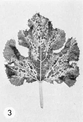
Diagnostic species
- Citrullus vulgaris
(watermelon). Systemic vein-banding, mosaic and leaf-distortion. - Cucurbita pepo (pumpkin). Interveinal chlorosis, mosaic, raised green blisters and leaf
distortion
(Fig. 1,
Fig. 2,
Fig. 3,
Fig. 4).
- Momordica balsamina. Immune.
-
Propagation species
- Cucurbita pepo
(pumpkin). Inoculate the cotyledons before emergence of the first true leaf and harvest after 3 weeks.Assay species
- Strains not restricted to cucurbits produce chlorotic and necrotic lesions in Chenopodium amaranticolor. All the isolates studied by Molnar & Schmelzer (1964) produced necrotic local lesions in Lavatera trimestris.
Strains
There are two major groups of strains, originally thought to belong to two separate viruses, WMV-1 and WMV-2 (Webb & Scott, 1965, see ‘Host Range and Symptomatology’ and ‘Relationships’). Many variants of the WMV-2 type have been described (Grogan et al., 1959; Van Regenmortel et al., 1962; Inouye, 1964; Webb & Scott, 1965; Milne & Grogan, 1969), but fewer of the WMV-1 type (Milne & Grogan, 1969).
Transmission by Vectors
Transmitted in the non-persistent manner by the aphids Myzus persicae, Aphis gossypii (Anderson, 1954), Aphis fabae (Molnar & Schmelzer, 1964) and several others (Coudriet, 1962). One strain not transmissible by aphids has been described (Molnar & Schmelzer, 1964).
Transmission through Seed
Lindberg et al. (1956) referred to one of their isolates (RMV) as being seed-borne. However, Grogan et al. (1959) found no seed transmission in Cucurbita maxima, C. pepo, Cucumis melo or Citrullus vulgaris, and they considered the seed-borne isolate of Rader, Fitzpatrick & Hildebrand (1947) to be a strain of squash mosaic virus.
Transmission by Dodder
Not tested.
Serology
Difficulties in obtaining reliable precipitin reactions with plant sap (Webb & Scott, 1965) may be due to loss of virus from clarified sap due to particle aggregation (Van Regenmortel, 1961). Antisera have been obtained with titres to virus of 1/512-1/16,000 but no detectable antibody to plant proteins (Van Regenmortel et al., 1962; Milne & Grogan, 1969). Intact virus does not react in gel-diffusion tests, but preparations degraded with 1% sodium dibutylnaphthalene sulphonate (Leonil SA) form specific precipitin bands in agar (Milne & Grogan, 1969). Serological detection of the virus in plants is unreliable.
Relationships
Isolates belonging to the WMV-1 and WMV-2 types, although originally classed by Webb & Scott (1965) as two separate viruses, according to their ability to infect non-cucurbitaceous species and on the basis of negative serological results, are now known to be serologically related (Milne & Grogan, 1969). They have similar particles and cannot be differentiated by cross-protection or cross-absorption serological tests (Milne & Grogan, 1969).
Watermelon mosaic virus is a member of the potato virus Y group of viruses, to some of which it is serologically related - closely to papaya ringspot virus (Milne & Grogan, 1969) and more distantly to potato Y and bean yellow mosaic viruses (Van Regenmortel et al., 1962).
Stability in Sap
In Cucurbita pepo sap the virus loses infectivity after 10 min at 58-65°C or 20-50 days at 20°C. Dilution end-point of sap from Cucurbita pepo is 10-4-10-5 and of sap from Citrullus vulgaris 10-2-10-4.
Purification
Virus particles aggregate readily and this may cause considerable losses during purification (Van Regenmortel, 1961). Homogenization of infected Cucurbita pepo leaves with citrate buffer, clarification with chloroform, differential centrifugation and zone electrophoresis in sucrose density gradients yields preparations that contain no detectable plant antigenic impurities (Van Regenmortel, 1964a, 1964 b). Good results have also been obtained by extracting with phosphate buffer, clarifying with 8.5% (v/v) n-butanol and zone electrophoresis (Milne & Grogan, 1969). Procedures involving DEAE chromatography, gel filtration and density gradient centrifugation were less successful (Van Regenmortel, 1961, 1964b; Milne & Grogan, 1969). Because of aggregation, infectivity is an unreliable guide to virus concentration; a hundredfold increase in infectivity end point was observed after ultrasonic treatment of aggregated preparations (Van Regenmortel, 1964a).
Properties of Particles
The particles precipitate in plant sap at pH 4.9 (Van Regenmortel, 1961) and the electrophoretic Rø value (Van Regenmortel, 1968, 1969) is 0.25.
Particle Structure
Particles are flexuous filaments (Fig. 5); reported values for the normal length range from 725 nm (Van Regenmortel et al., 1962) to 746-765 nm (Schmelzer, 1966; Purcifull, Edwardson & Christie, 1968; Milne & Grogan, 1969)
Particle Composition
No reports.
Relations with Cells and Tissues
Striated inclusions and ‘pinwheels’ have been reported in host cells (Edwardson, 1966; Purcifull, 1968; Purcifull et al., 1968).
Notes
Considerable confusion and synonymity among allegedly separate cucurbit viruses arose from distinctions based solely on host reactions (Grogan et al., 1959). The virus can be distinguished from squash mosaic and cucumber mosaic viruses because it infects Citrullus vulgaris systemically and can be separated from cucumber green mottle mosaic and tobacco ringspot viruses because it is transmitted by aphids.
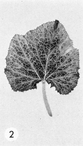
Figures

Healthy leaf of Cucurbita pepo.
Interveinal chlorosis in C. pepo.
Raised green blisters in C. pepo.

Leaf distortion in C. pepo.

Virus particles, shadowed, prepared by dip method. Bar represents 1 µm.
References list for DPV: Watermelon mosaic virus (63)
- Anderson, Phytopathology 44: 198, 1954.
- Coudriet, J. econ. Ent. 55: 519, 1962.
- Edwardson, Am. J. Bot. 53: 359, 1966.
- Grogan, Hall & Kimble, Phytopathology 49: 366, 1959.
- Inouye, Ber. Ohara Inst. landw. Biol. 12: 133, 1964.
- Komuro, Ann. phytopath. Soc. Japan 22: 220, 1957.
- Lindberg, Hall & Walker, Phytopathology 46: 489, 1956.
- Milne & Grogan, Phytopathology 59: 809, 1969.
- Molnar & Schmelzer, Phytopath. Z. 51: 361, 1964.
- Purcifull, Virology 36: 690, 1968.
- Purcifull, Edwardson & Christie, Virology 35: 478, 1968.
- Rader, Fitzpatrick & Hildebrand, Phytopathology 37: 809, 1947.
- Schmelzer, Naturwissenschaften 53: 619, 1966.
- Schmelzer & Milicic, Phytopath. Z. 57: 8,1966.
- Toba, Pl. Dis. Reptr 46: 409, 1962.
- Van Regenmortel, Virology 12: 127, 1960.
- Van Regenmortel, S. Afr. J. agric. Sci. 4: 405, 1961.
- Van Regenmortel, Virology 23: 495, 1964a.
- Van Regenmortel, Phytopathology 54: 282, 1964b.
- Van Regenmortel, S. Afr med. J. 42: 118, 1968.
- Van Regenmortel, Annls Phytopath. 1: 127, 1969.
- Van Regenmortel, Brandes & Bercks, Phytopath. Z. 45: 205, 1962.
- Webb & Scott, Phytopathology 55: 895, 1965.